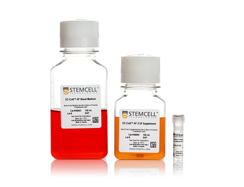
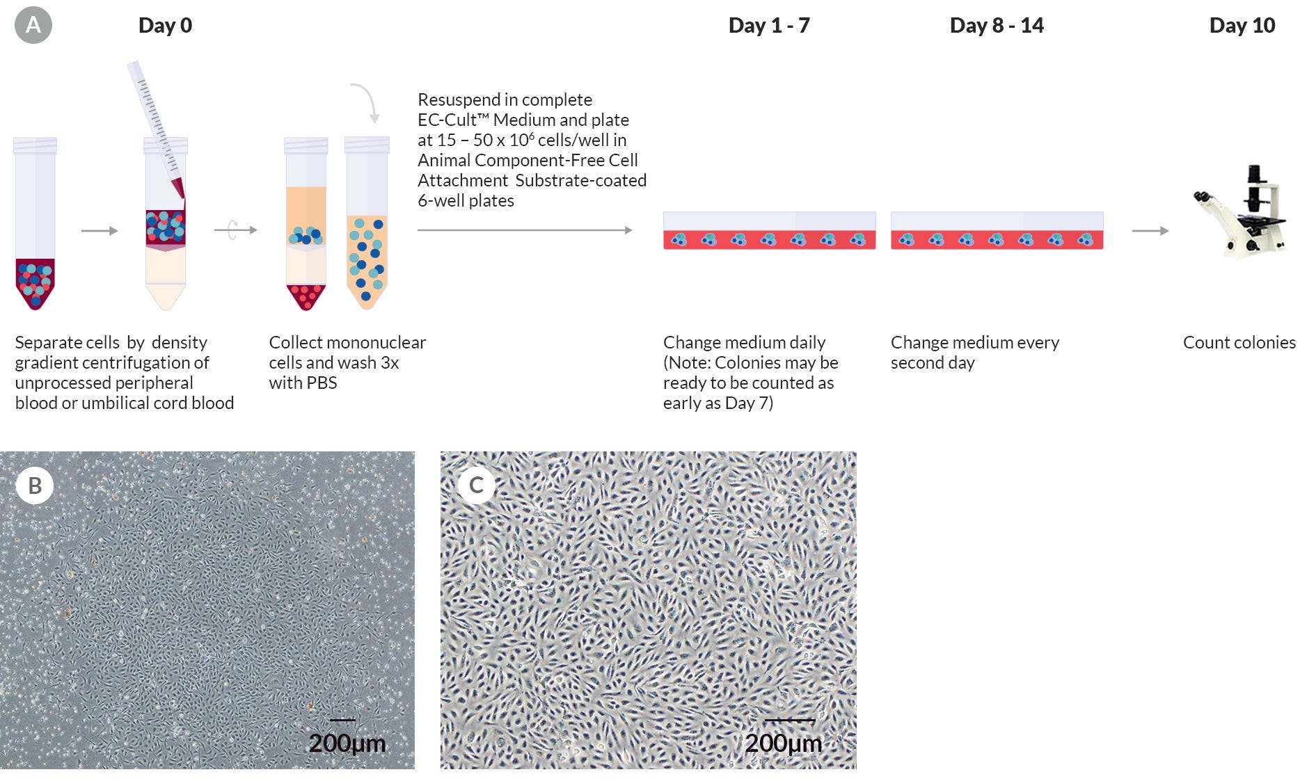
EC-Cult™-XF Culture Kit
Culture kit for derivation and proliferation of human endothelial colony-forming cells (ECFCs) and mature endothelial cells
Request Pricing
Thank you for your interest in this product. Please provide us with your contact information and your local representative will contact you with a customized quote. Where appropriate, they can also assist you with a(n):
Estimated delivery time for your area
Product sample or exclusive offer
In-lab demonstration
-
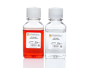 Animal Component-Free Cell Dissociation Kit
Dissociation kit for human stem and progenitor cells
-
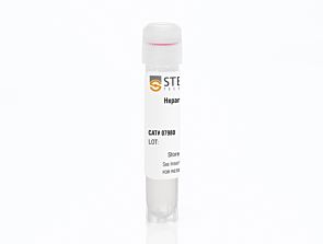 Heparin Solution
Cell culture supplement
-
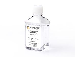 D-PBS (Without Ca++ and Mg++)
Dulbecco’s phosphate-buffered saline without calcium and magnesium
Overview
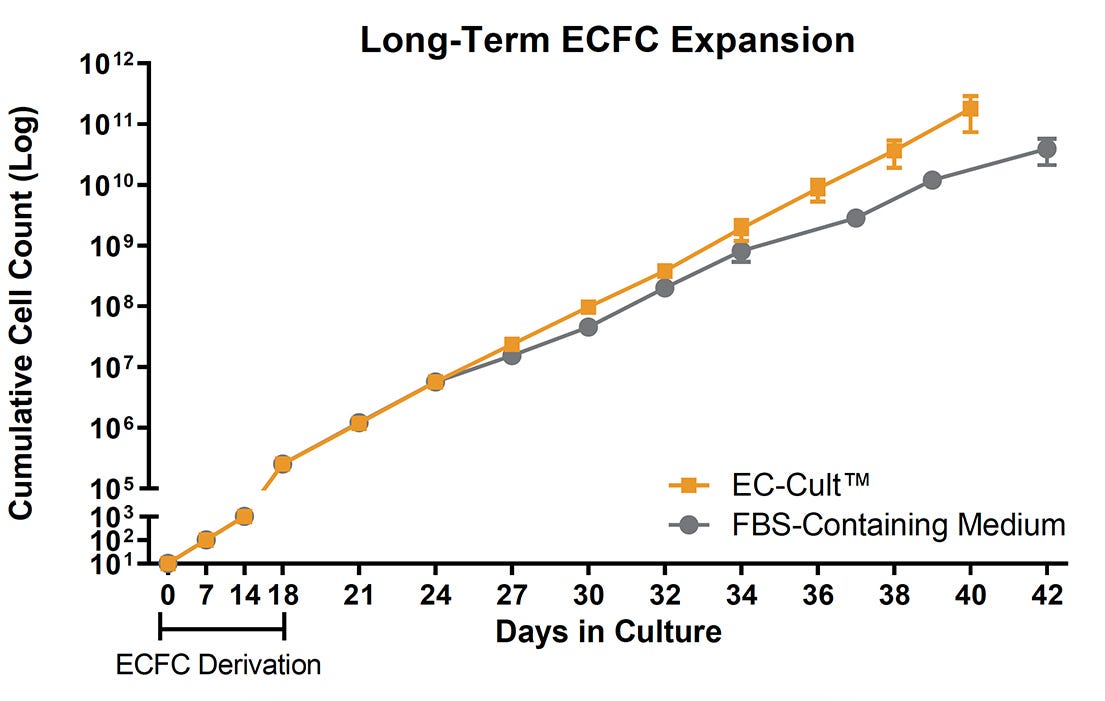
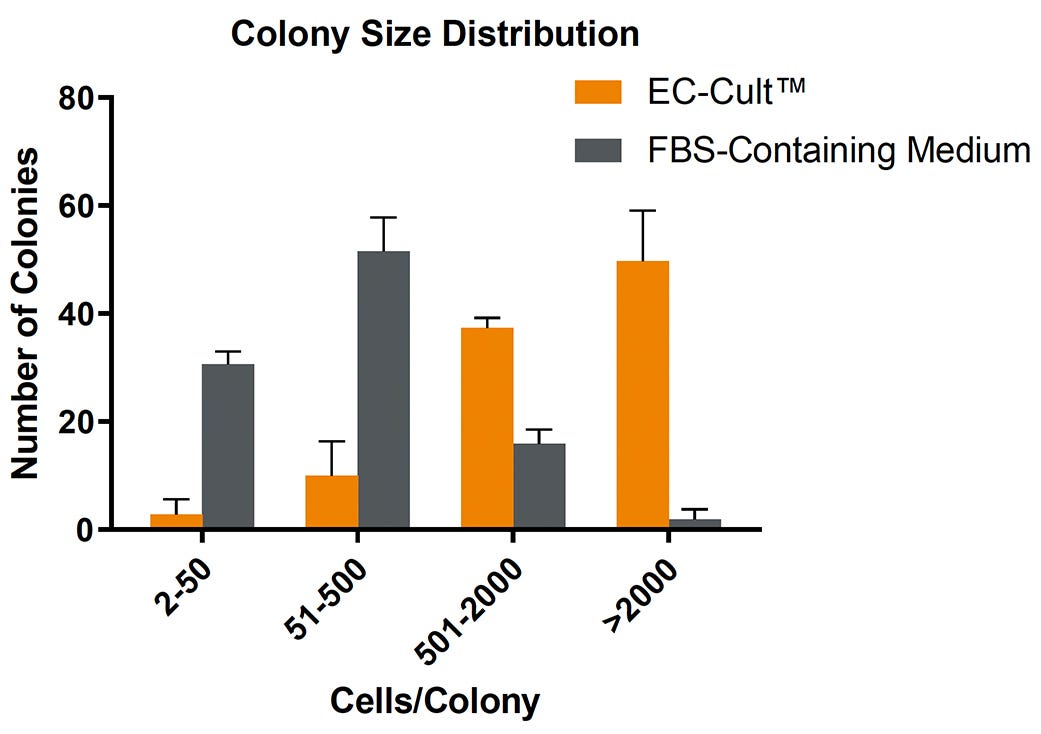
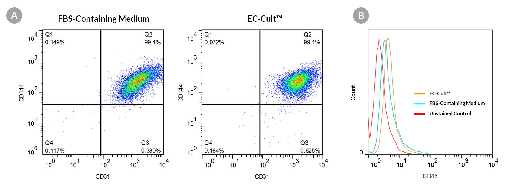
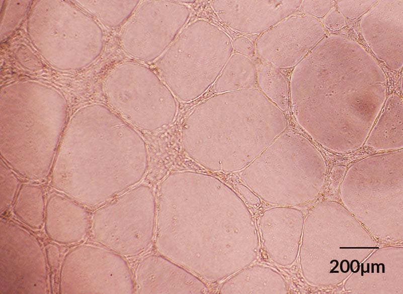
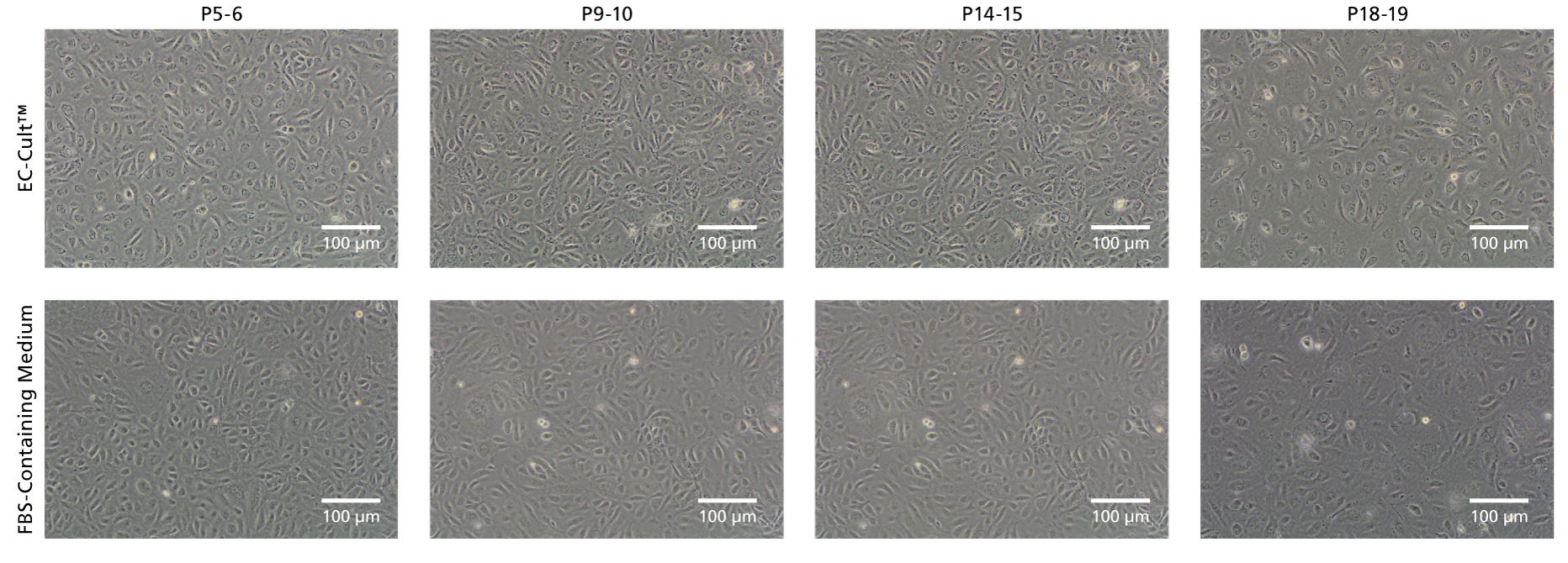
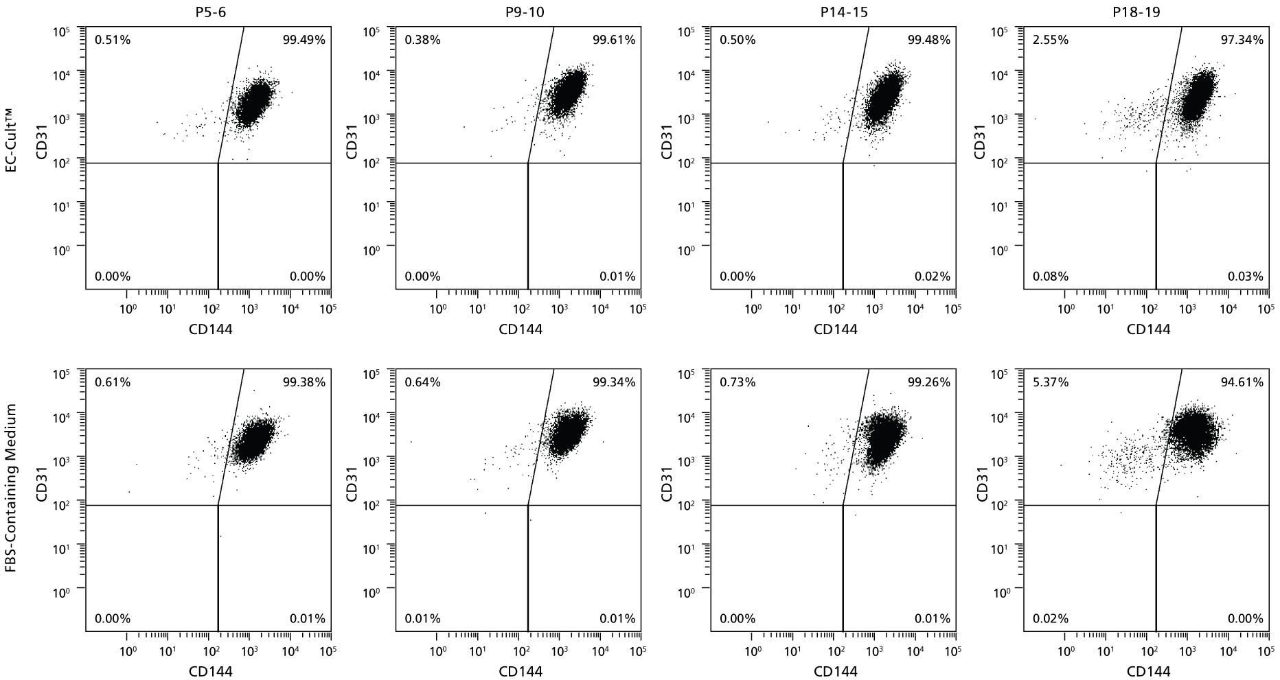
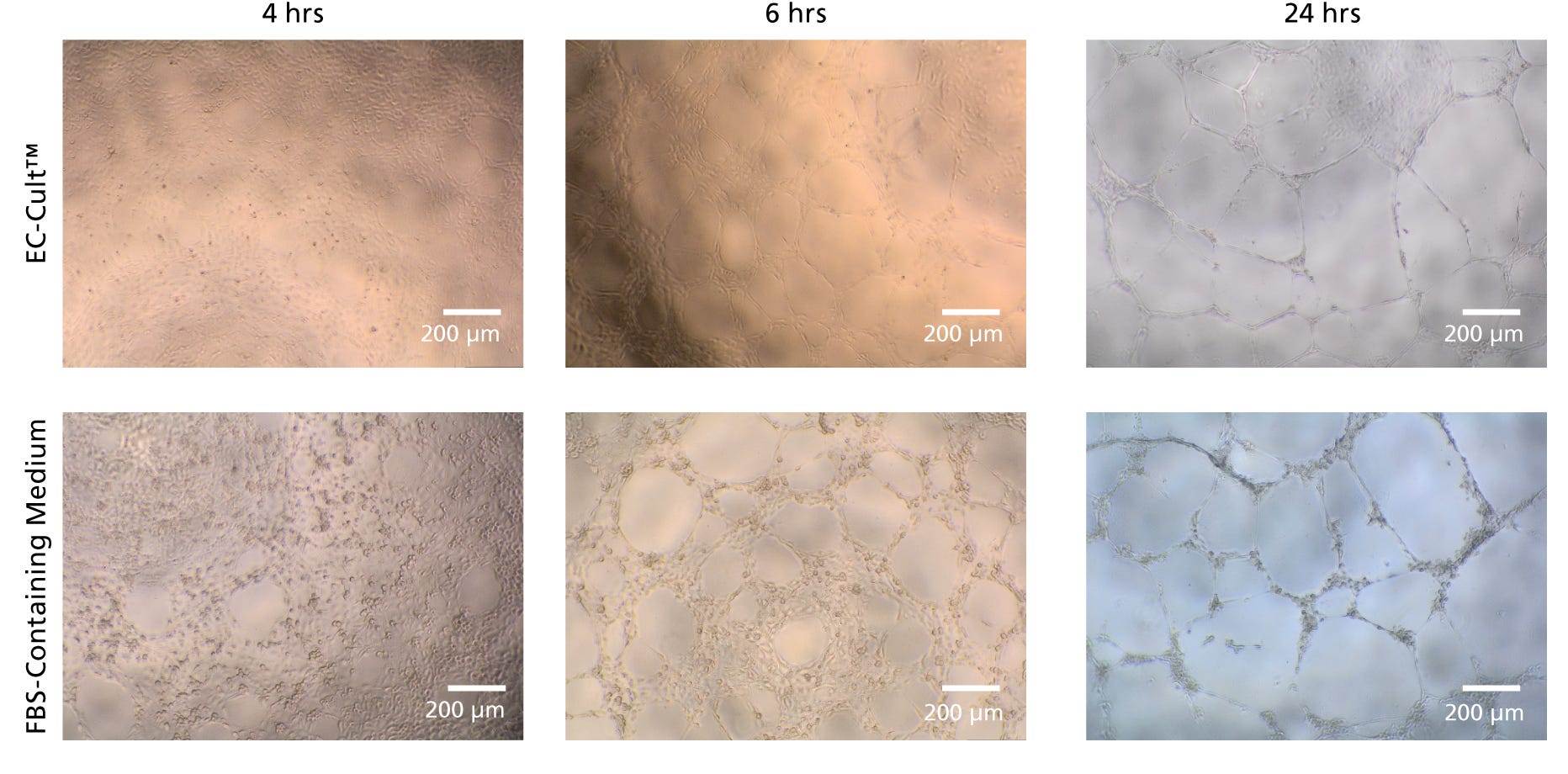
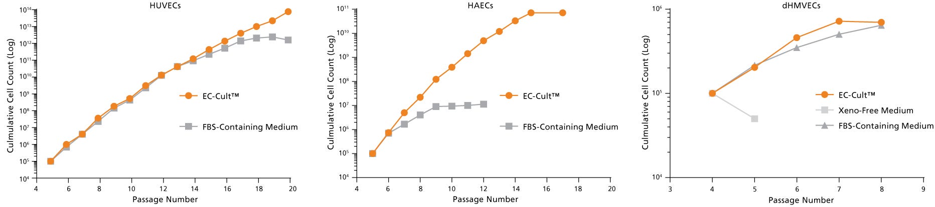
Data Figures
Protocols and Documentation
Find supporting information and directions for use in the Product Information Sheet or explore additional protocols below.
Resources and Publications
Educational Materials (4)


Publications (2)
Abstract
Abstract
Related Products

Legal Statement:
The included rhCollagen is supplied by
Quality Statement:
PRODUCTS ARE FOR RESEARCH USE ONLY AND NOT INTENDED FOR HUMAN OR ANIMAL DIAGNOSTIC OR THERAPEUTIC USES UNLESS OTHERWISE STATED. FOR ADDITIONAL INFORMATION ON QUALITY AT STEMCELL, REFER TO WWW.STEMCELL.COM/COMPLIANCE.
