CELLTREAT® 0.4 µm Polyethylene Membrane Inserts

Polystyrene plate with lid and polyethylene membrane inserts

CELLTREAT® 0.4 µm Polyethylene Membrane Inserts

Polystyrene plate with lid and polyethylene membrane inserts

From: 81 USD
Catalog #
(Select a product)
Polystyrene plate with lid and polyethylene membrane inserts
Add to Wish List

What's Included

  • CELLTREAT® 0.4 µm Polyethylene Membrane Inserts
    • 24-Well Format (Catalog #100-0997)
      • 2 x 24-Well Plates with 12 Inserts/Plate
      • Alternative to: Costar® Catalog #38024
    • 12-Well Format (Catalog #100-1027)
      • 2 x 12-Well Plates with 12 Inserts/Plate
      • Alternative to: Costar® Catalog #38023
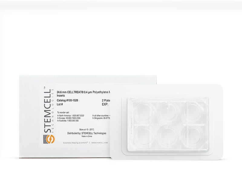
    • 6-Well Format (Catalog #100-1026)
      • 2 x 6-Well Plates with 6 Inserts/Plate

Overview

Culture airway epithelial cells at the air-liquid interface with CELLTREAT® 0.4 µm Polyethylene Membrane Inserts. These inserts support the growth of both anchorage-dependent and anchorage-independent cells, including those that are fed at the basolateral surface or in co-culture systems. With a pore density of 2 x 10⁶ pores/cm², these permeable inserts are useful for a variety of applications, including infectious and genetic disease modeling, drug discovery, toxicity testing, and epithelial cell biology. CELLTREAT® 0.4 µm Polyethylene Membrane Inserts are verified for use with PneumaCult™-Ex Plus Medium (Catalog #05040), PneumaCult™-ALI Medium (Catalog #05001), and IntestiCult™ Organoid Differentiation Medium (Human; Catalog #100-0214).
Subtype
Dishes and Plates
Species
Human, Mouse, Non-Human Primate, Other, Rat
Application
Cell Culture
Brand
CELLTREAT

Data Figures

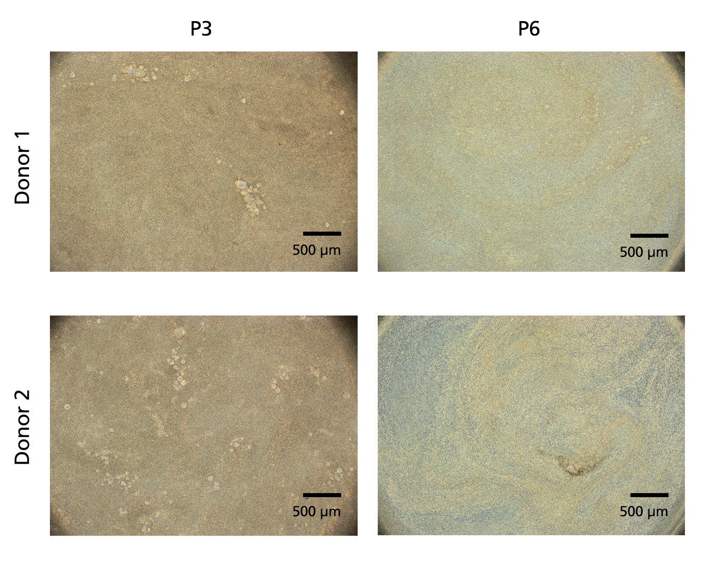
Figure 1. ALI Cultures Generated Using CELLTREATⓇ Inserts Are Highly Differentiated

Human bronchial epithelial cells (HBECs), expanded in PneumaCult™ Ex-Plus Medium (Catalog #05040), were seeded onto CELLTREATⓇ Inserts at an early (P3) or late passage (P6) and differentiated in PneumaCult™-ALI Medium (Catalog #05001) for 4 weeks. A high level of differentiation (represented by brown areas of the culture), as well as mucus secretion and visible cilia beating on the apical surface (not shown), were observed with cultures grown on CELLTREATⓇ Inserts. Scale bar = 500 µm.

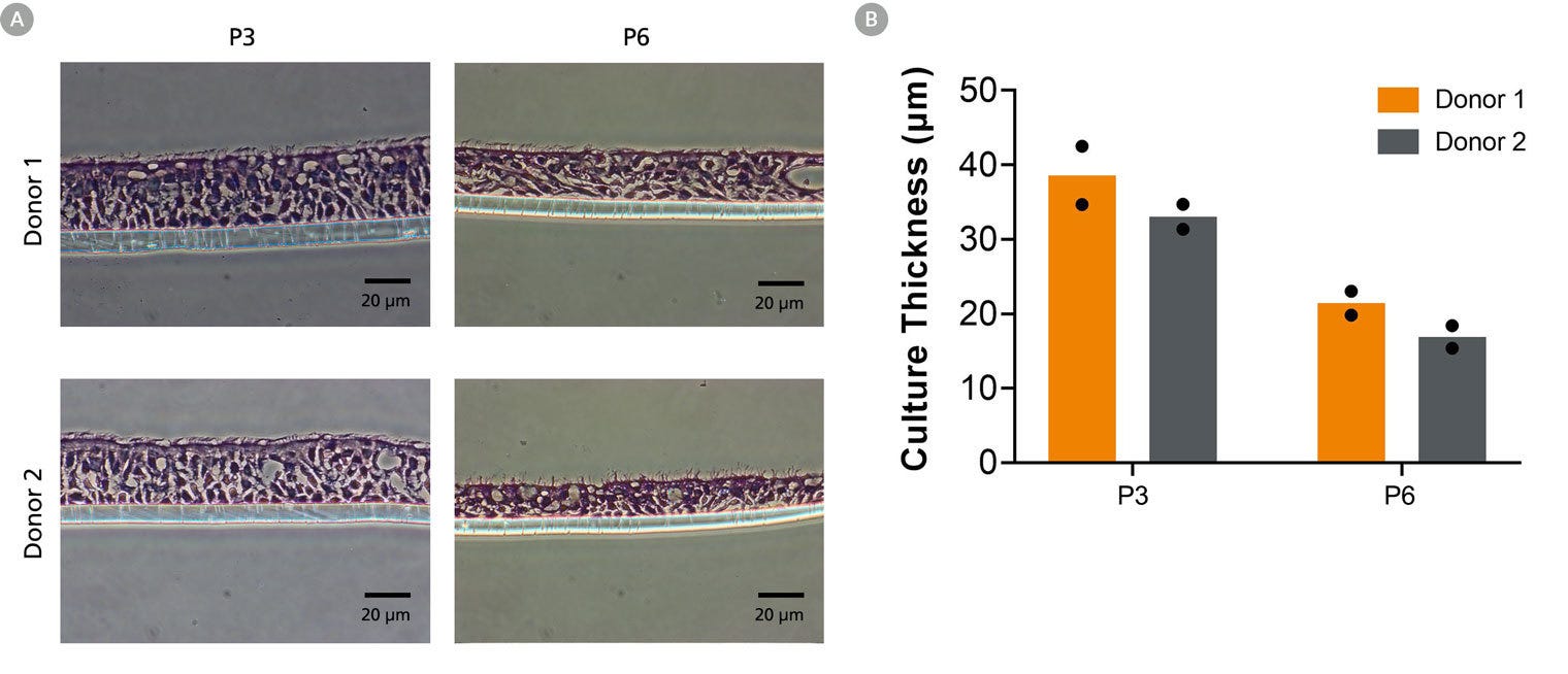
Figure 2. ALI Cultures Generated Using CELLTREATⓇ Inserts Form Pseudostratified Columnar Epithelium

ALI cultures, differentiated from HBECs at an early (P3) or late passage (P6), were fixed 4 weeks after air-lift. They were then paraffin embedded, sectioned, and stained with H&E. Cultures generated using CELLTREATⓇ Inserts resulted in a pseudostratified columnar epithelium with visible cilia at the apical surface. An expected reduction in ALI culture thickness is observed with increasing HBEC passage number. (A) Representative H&E stained cross-sections; (B) ALI culture thickness (n = 2 replicate wells from a single experiment). Scale bar = 20 µm.

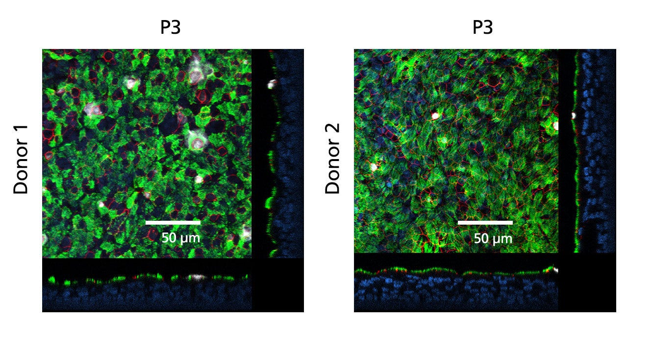
Figure 3. ALI Cultures Generated Using CELLTREATⓇ Inserts Enable Differentiation of HBECs to Cell Types Present in the Large Airway Epithelium

ALI cultures differentiated from HBECs at an early passage (P3) were fixed and stained with antibodies for ciliated cells (AC-tubulin; green), cell junctions (ZO-1; red), and goblet cells (Muc5AC; white). The nuclei were counterstained with DAPI (blue). Pseudostratified mucociliary differentiation is indicated by high expression of markers of the large airway. Scale bar = 50 µm.

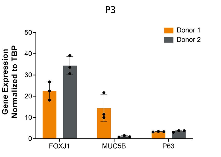
Figure 4. ALI Cultures Generated Using CELLTREATⓇ Inserts Show High Expression of Large Airway Markers

ALI cultures, differentiated from HBECs at an early passage (P3), were collected 4 weeks after air-lift. Expression of large airway markers for ciliated (FOXJ1), goblet (MUC5B), and basal (P63) cells were assessed by qPCR and normalized to the housekeeping gene TBP. Error bars represent standard deviation (n = 3 replicate wells from a single experiment).

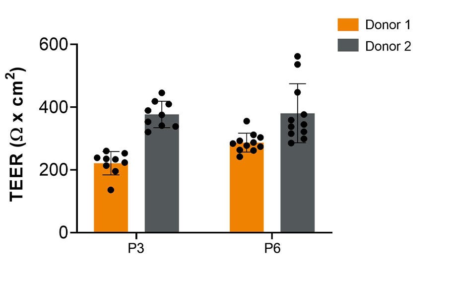
Figure 5. ALI Cultures Generated Using CELLTREATⓇ Inserts Show Optimal Barrier Function

TEER measurements of ALI cultures differentiated from HBECs at an early (P3) or late passage (P6) were taken 4 weeks after air-lift. Values were corrected against blank wells. Average values of donors were within the expected normal physiological levels (200 - 800 Ω x cm2), indicative of optimal culture differentiation. Error bars represent standard deviation (n ≥ 9 replicate wells from a single experiment).

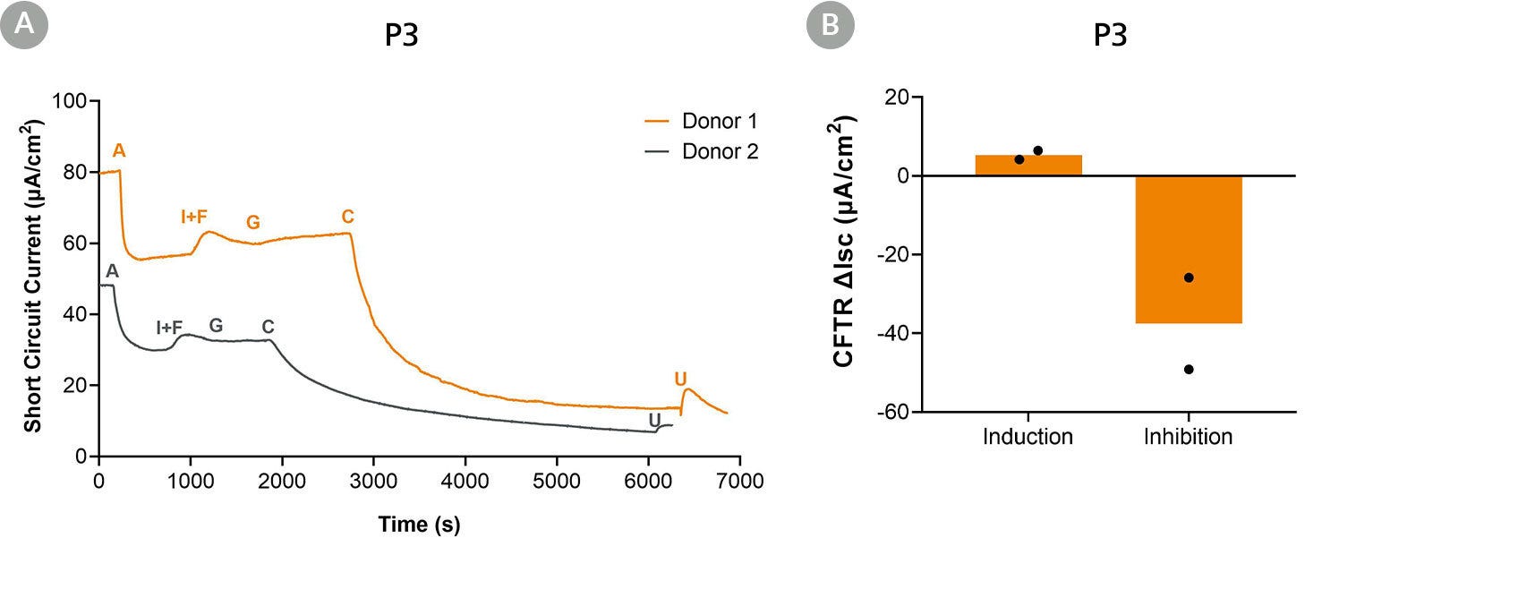
Figure 6. ALI Cultures Generated Using CELLTREATⓇ Inserts Show Normal Electrophysiological Activity

ALI cultures differentiated from HBECs at an early passage (P3) were assessed using Ussing Chamber analysis 6 months after air-lift. Cultures generated using CELLTREATⓇ Inserts showed functional ion-transporters within the epithelial cell layer, as demonstrated by the addition of activating and inhibitory drugs. (A) Representative electrophysiological trace of two independent donors; (B) CFTR-inducible and inhibitable short-circuit current (n = 2 donors). A = Amiloride (ENaC inhibitor); I+F = IBMX and Forskolin (CFTR activators); G = Genistein (CFTR potentiator); C = CFTRinh-172 (CFTR inhibitor); U = UTP (calcium-activated chloride channels (CaCCs) activator).

Figure 7. Differentiated Intestinal Organoid-Derived Monolayers or Caco-2 Cells Reach Full Confluence When Using CELLTREATⓇ Inserts

Human intestinal organoids, previously maintained in IntestiCult™ Organoid Growth Medium (Human) (Catalog #06010), were dissociated, replated onto CELLTREATⓇ Inserts, and differentiated using IntestiCult™ Organoid Differentiation Medium (Human) (Catalog #100-0214). The Caco-2 epithelial cell line from a colorectal adenocarcinoma was used as a control. Between 3 to 7 days of culture, monolayers from two different ileal donors and Caco-2 cells reached full confluence. Scale bar = 250 µm.

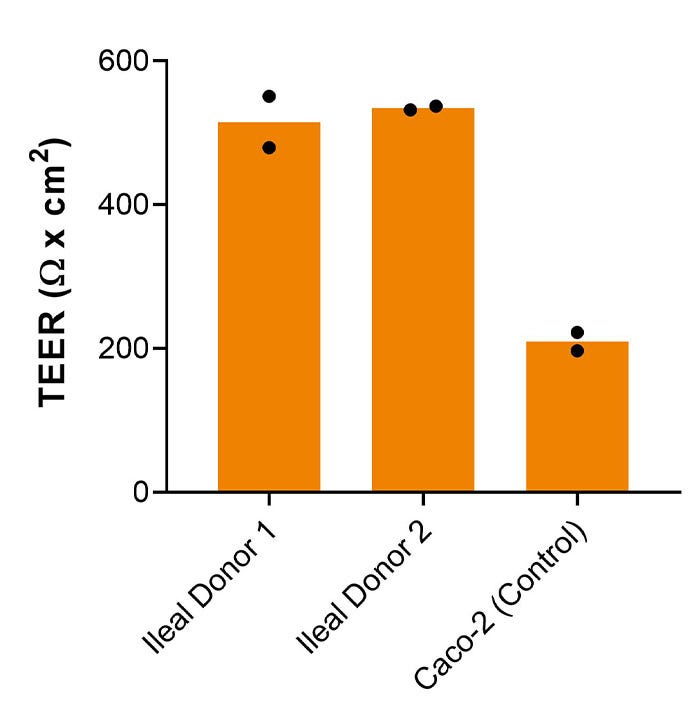
Figure 8. Differentiated Intestinal Organoid Derived Monolayers or Caco-2 Cells Generate Using CELLTREATⓇ Inserts Demonstrate Optimal Barrier Function

TEER measurements of human intestinal organoid monolayers or the epithelial cell line, Caco-2, were taken 7 days after plating. Cultures generated using CELLTREATⓇ Inserts demonstrate values within the expected range of over 200 Ω x cm2, indicative of optimal barrier function (n = 2 replicate wells from a single experiment).

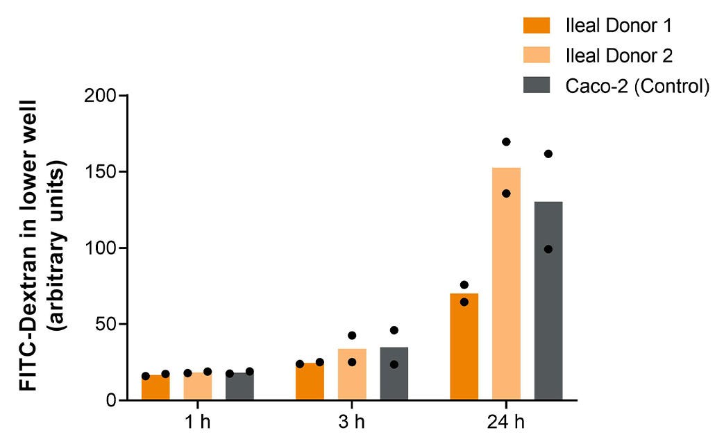
Figure 9. Differentiated Intestinal Organoid Derived Monolayers or Caco-2 Cells Generated Using CELLTREATⓇ Inserts Show Low Permeability to FITC-Dextran

The permeability of human intestinal organoid monolayers or the epithelial cell line, Caco-2, were measured 7 days after plating. FITC-dextran was added apically and sampled from the basolateral chamber after 1, 3, and 24 h. All cultures generated using CELLTREATⓇ Inserts demonstrate low permeability with the steady diffusion of FITC-dextran observed over time, indicative of a confluent monolayer (n = 2 replicate wells from a single experiment).

Protocols and Documentation

Find supporting information and directions for use in the Product Information Sheet or explore additional protocols below.

Document Type
Product Name
Catalog #
Lot #
Language
Catalog #
100-1027
Lot #
All
Language
English
Catalog #
100-1026
Lot #
All
Language
English
Catalog #
100-0997
Lot #
All
Language
English

Applications

This product is designed for use in the following research area(s) as part of the highlighted workflow stage(s). Explore these workflows to learn more about the other products we offer to support each research area.