OptiPrep™

Density gradient medium for the isolation of viruses, organelles, macromolecules, or cells
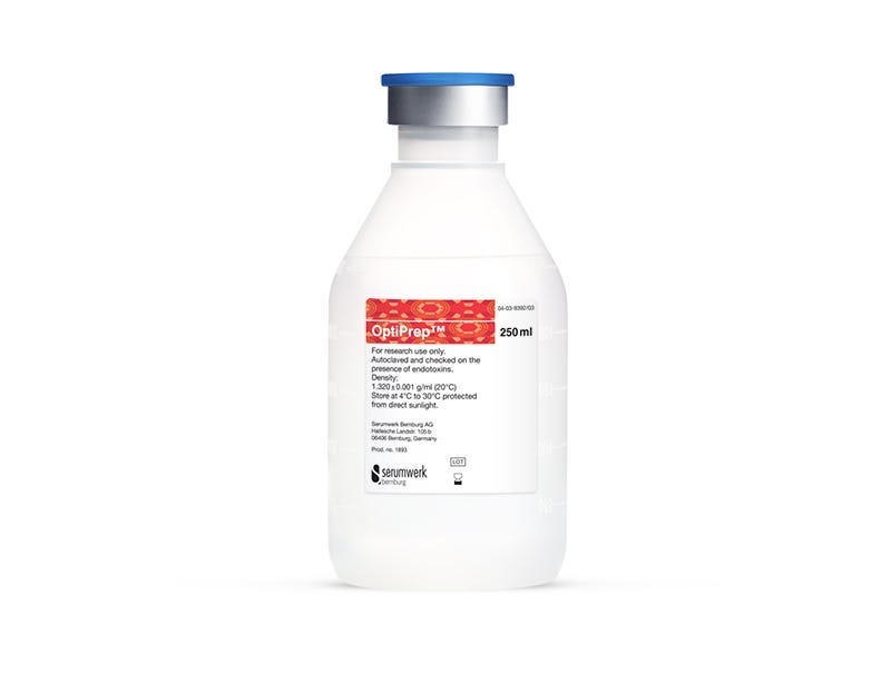
OptiPrep™

Density gradient medium for the isolation of viruses, organelles, macromolecules, or cells

From: 289 USD
Catalog #
(Select a product)
Density gradient medium for the isolation of viruses, organelles, macromolecules, or cells
Add to Wish List

Product Advantages


  • Flexible: Use to form continuous or discontinuous gradients

  • Versatile: Customize separation characteristics for isolation of viruses, organelles, macromolecules, or cells

  • Gentle: Non-ionic, non-toxic to cells, isoosmotic, and metabolically inert

Overview

Simplify the isolation and purification of macromolecules; viruses; a wide range of cell types; and organelles such as nuclei, mitochondria, endosomes, or exosomes by using OptiPrep™ medium. This flexible, versatile, and gentle density gradient medium is non-ionic, iodixanol-based (60% w/v) and has a density of 1.320 ± 0.001 g/mL.
Contains
• Iodixanol: 60% (w/v)
• Density: 1.320 ± 0.001 g/mL
Cell Type
Mononuclear Cells, Other
Species
Human, Mouse, Non-Human Primate, Other, Rat
Application
Cell Isolation
Brand
OptiPrep
Area of Interest
Immunology

Protocols and Documentation

Find supporting information and directions for use in the Product Information Sheet or explore additional protocols below.

Document Type
Product Name
Catalog #
Lot #
Language
Product Name
OptiPrep™
Catalog #
07820
Lot #
All
Language
English
Document Type
Safety Data Sheet
Product Name
OptiPrep™
Catalog #
07820
Lot #
All
Language
English

Applications

This product is designed for use in the following research area(s) as part of the highlighted workflow stage(s). Explore these workflows to learn more about the other products we offer to support each research area.

Resources and Publications

Educational Materials (1)

Publications (19)

sc-rDSeq: Droplet-based single-cell full-length total RNA-seq method. X. Sun and O. Ram Biology methods & protocols 2026 May

Abstract

This protocol describes sc‑rDSeq, a scalable, droplet‑based method for full‑length, strand‑specific total RNA sequencing at single‑cell resolution. The protocol uses a refined set of 220 ribosomal‑depleted sequences (rDS) primers that selectively exclude ribosomal RNA during initial reverse transcription, enabling capture of both polyadenylated and non‑polyadenylated RNAs such as histone RNAs, noncoding RNAs, and enhancer RNAs, without requiring costly post‑amplification depletion steps. This method is useful for researchers who would like to detect not only gene expression variations, but also alternative splicing events and single nucleotide variations in complex heterogenous cellular systems, providing a more complete view of cellular heterogeneity and regulatory programs that remain invisible to conventional polyadenylated‑only sequencing approaches. Compared with existing full‑length protocols, which are often limited by high reagent costs or reliance on complex multistep microfluidics, sc-rDSeq provides a simpler, single-step microfluidic workflow compatible with standard inDrops platforms, which may reduce experimental complexity and cost relative to existing full-length total-RNA methods. A key improvement is the 10-fold increase in unique molecular identifiers per cell relative to 3' end‑based methods, at a reported reagent cost of approximately $0.08 per cell, making deep total transcriptome analysis more accessible. The protocol includes three major parts: sc‑rDSeq barcode synthesis, single‑cell co‑encapsulation, and library construction.
Rapid Volumetric Bioprinting Coupled with Dynamic Perfusion Enhances Human Hepatic Organoid Toxicity Testing. Y. Tao et al. Cells 2026 Jul

Abstract

Drug-induced liver injury (DILI) remains a major cause of acute liver failure and drug withdrawal from the market. Recently developed three-dimensional (3D) hepatic in vitro systems exhibit improved functionality and drug sensitivity compared with conventional two-dimensional cultures. These 3D models range from simple physiologic-like culture systems to advanced bioreactors with dynamic flow to provide sufficient nutrients and consistent drug exposure. However, whether dynamic perfusion improves sensitivity and reproducibility of hepatotoxicity testing remains unclear. Here, we developed a tailor-made perfusion platform to support volumetric bioprinted hepatic constructs for hepatotoxicity testing. The constructs consist of intrahepatic cholangiocyte organoids (ICOs) differentiated towards hepatocyte lineage and embedded in a gelatin methacryloyl bioresin. For toxicity evaluation, the hepatocyte-like ICO constructs were exposed to prolonged subtoxic acetaminophen treatment (10 mM, 7 days). The perfusion system effectively maintained and enhanced hepatocyte differentiation, evidenced by upregulated hepatic markers under perfused conditions compared to static controls. Testing of acetaminophen hepatotoxicity revealed that the perfused constructs displayed elevated cellular injury, with markedly higher liver injury markers relative to controls. Collectively, this study demonstrates the successful application of perfusion-based 3D model culture and highlights its potential as a more physiological platform for hepatotoxicity risk assessment in drug discovery and regenerative medicine.
Multicompartment hydrogel microcapsules for creating spatially patterned cell co-cultures S. Cho et al. Microsystems & Nanoengineering 2026 Jan

Abstract

There is increasing clinical evidence that pancreatic dysfunction in diabetes needs to be viewed in the context of crosstalk with the liver as well as other organs. Our goal for this study was to develop a pancreas-liver co-culture system suited for mechanistic and therapy testing studies in the context of multi-organ cross talk. To achieve this goal, we developed a co-axial flow-focusing microfluidic device to fabricate multi-compartment hydrogel microcapsules. Each microcapsule contained two aqueous compartments or cores surrounded by poly(ethylene glycol) (PEG) hydrogel. Each microcapsule had pancreatic β-cells loaded into one compartment and hepatic cells into another compartment. Individual encapsulated cells assembled into pancreatic and hepatic cell spheroids over time. Characterization of microcapsules revealed enhanced hepatic and pancreatic function in microcapsules containing pancreas-liver co-cultures compared to microcapsules with one cell type only. Multicompartment microcapsules represent a novel microphysiological system type and hold the promise of increasing experiment throughput for mechanism discovery and drug development studies.