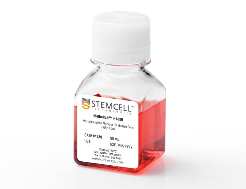
MethoCult™ H4330

Methylcellulose-based medium with EPO for human cells

MethoCult™ H4330

Methylcellulose-based medium with EPO for human cells
From: 651 USD
Catalog #
(Select a product)
Methylcellulose-based medium with EPO for human cells
Add to Wish List

Overview

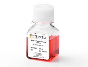
MethoCult™ H4330 is a methylcellulose-based medium for the growth and enumeration of hematopoietic progenitor cells in colony-forming unit (CFU) assays of human bone marrow, mobilized peripheral blood, peripheral blood, and cord blood samples. This formulation contains erythropoietin (EPO), but does not contain other cytokines. It is recommended for assays of late erythroid progenitors, specifically colony-forming cell - erythroid (CFU-E), in the presence of EPO only. It is also recommended for assays of early erythroid progenitors, specifically burst-forming unit - erythroid (BFU-E), and other colony-forming cells when appropriate cytokines are added. It can also be used to assess colony-stimulating factor activity or burst-promoting activity in unknown samples.

Browse our Frequently Asked Questions (FAQs) on performing the CFU assay and explore its utility as part of the cell therapy workflow.
Contains
• Methylcellulose in Iscove's MDM
• Fetal bovine serum
• Bovine serum albumin
• 2-Mercaptoethanol
• Recombinant human erythropoietin (EPO)
• Supplements
Subtype
Semi-Solid Media, Specialized Media
Cell Type
Hematopoietic Stem and Progenitor Cells
Species
Human, Non-Human Primate
Application
Cell Culture, Colony Assay, Functional Assay
Brand
MethoCult
Area of Interest
Drug Discovery and Toxicity Testing, Stem Cell Biology

Protocols and Documentation

Find supporting information and directions for use in the Product Information Sheet or explore additional protocols below.

Document Type
Product Name
Catalog #
Lot #
Language
Catalog #
04330
Lot #
All
Language
English
Document Type
Technical Manual
Catalog #
04330
Lot #
All
Language
English
Document Type
Safety Data Sheet
Catalog #
04330
Lot #
All
Language
English

Applications

This product is designed for use in the following research area(s) as part of the highlighted workflow stage(s). Explore these workflows to learn more about the other products we offer to support each research area.

Resources and Publications

Frequently Asked Questions

Why use semi-solid media?

Semi-solid media (methylcellulose-based MethoCult™ and collagen-based MegaCult™-C) allow the clonal progeny of a single progenitor cell to remain spatially isolated from other colonies within a culture, so they may be separately identified and counted.

Why use methylcellulose-based media?

Methylcellulose permits better growth of erythroid colonies than other types of semi-solid support systems (eg. agar) while allowing optimal myeloid colony formation. When appropriate cytokines are present, committed progenitor cells of both erythroid and granulocyte/macrophage lineages (CFU-GM, CFU-G, CFU-M) as well as multi-potential progenitor cells (CFU-GEMM), can be assayed simultaneously in the same culture dish.

Is it necessary to add antibiotics to the media?

No, aseptic technique should be sufficient to maintain sterile cultures. However, antibiotics (eg. Penicillin/Streptomycin) or anti-fungals (eg. Amphotericin B) may be added to the methylcellulose medium if desired.

Is there anything I can do if my cultures appear contaminated?

No, once contamination is visible, it is not possible to rescue the cultures by the addition of antibiotics. Bacteria and yeast inhibit colony formation by depleting nutrients or by releasing toxic substances.

Why can't I use a pipette to dispense methylcellulose-based media?

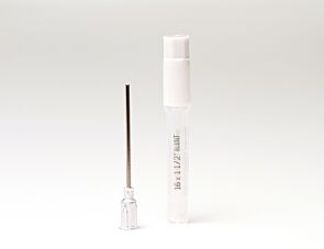
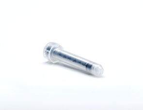
Methylcellulose is a viscous solution that cannot be accurately dispensed using a pipette due to adherence of the medium to the walls of the pipette tip. Blunt-End, 16 Gauge needles (Catalog #28110), in combination with 3 cc Syringes (Catalog #28230) are recommended for accurate dispensing of MethoCult™.

Can I 'pluck' the colonies for individual analysis?

Yes, colonies can be 'plucked' using a pipette with 200 µL sterile pipette tips or using a glass Pasteur pipette with an elongated tip. Individual colonies should be placed in a volume of 25 - 50 µL of medium, and diluted into suitable culture medium for further culture or analysis.

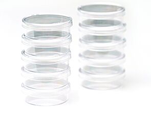
Why are low adherence dishes so important?

Adherent cells such as fibroblasts can cause inhibition of colony growth and obscure visualization of colonies.

Can MethoCult™ products be used for lymphoid progenitor CFU assays?

Human lymphoid progenitors (B, NK and T) seem to require stromal support for growth therefore cannot be grown in MethoCult™. Mouse pre-B clonogenic progenitors can be grown in MethoCult™ M3630 (Catalog #03630).

Is it possible to set up CFU assays in a 24-well plate?

Yes, as long as a plating concentration optimized for the smaller surface area of a well in a 24-well plate (1.9 cm2 as compared to ~9.5 cm2 for a 35 mm dish) is used for these assays. The number of replicate wells required to get an accurate estimation of CFU numbers may also need to be increased.

Can I stain colonies in MethoCult™ medium?

The cells in individual colonies in MethoCult™ can be stained, eg., for analysis of morphology or phenotype, after they are plucked from the dish and washed free of methylcellulose. Colonies grown in collagen-based MegaCult™-C medium can be used for immunohistochemical or enzymatic staining in situ after dehydration and fixation onto glass slides.

Are there differences in colony morphology with serum-free media?

Serum-containing media generally give better overall growth (colonies may appear larger) but there are no large differences in total colony numbers when CFU assays using serum-free media and serum-containing media are compared, provided that identical cytokines are present.

Can MethoCult™ be made with alternate base media?

Yes, this can be done as a 'custom' media order. Please contact techsupport@stemcell.com for more information.

Is there a MethoCult™ formulation suitable for HPP-CFC (high proliferative potential colony forming cell)?

Yes, MethoCult™ H4535 (Catalog #04535) can be used for the HPP-CFC assay as it does not contain EPO. The culture period is usually 28 days. It is not necessary to feed these cultures as growth factors in the medium are present in excess. As HPP-CFCs can be quite large, overplating can be a problem. It is recommended to plate cells at two or more different concentrations.

Publications (15)

Spatial transcriptomic analyses highlight distinct erythroid niches in mice and humans. X. Han et al. Nature genetics 2026 Jul

Abstract

Erythroid cells require specialized microenvironments called erythroblastic islands (EBIs), niches comprising a central macrophage surrounded by developing erythroid precursors, to complete their maturation. Understanding EBI composition and function has been limited by two-dimensional in vitro models and the unclear composition of EBIs in human hematopoietic tissues. Using spatial transcriptomic mapping in mouse and human hematopoietic tissues during development and under stress conditions, we show that EBI architecture is unexpectedly species-specific. In mice, C1q-expressing macrophages serve as a hallmark of EBI central macrophages and mediate clearance of ejected erythroid nuclei. In humans, however, EBIs are characterized by macrophage-independent erythroid clusters in fetal liver and bone marrow, whose integrity depends critically on the adhesion molecule ICAM4. These human erythroid clusters are disrupted in myeloid diseases but can be restored with therapy. These findings redefine conventional models of erythroid niche biology and establish a framework for understanding niche dynamics across species.
Development and Characterization of a Novel Congenital Acute Erythroid Leukemia Cell Line with Unique Features P. Sitaula et al. Cancers 2026 Apr

Abstract

Background: Acute erythroid leukemia (AEL) or AML-M6 predominantly affects older adults and is rare in childhood. Compared with other AML subtypes, AEL remains relatively understudied because of its rarity. We established LS-CHM, a novel AEL cell line derived from the ascitic fluid of a patient with congenital leukemia. Interestingly, leukemic cells persisted in the ascitic fluid even after successful eradication from the bone marrow and extramedullary sites. Method: Leukemia cells from the ascites fluid exhibited robust proliferation in culture independent of cytokine requirement and were further characterized by flow cytometric immunophenotyping, cytogenetics, cell cycle and doubling time analysis, colony formation, genome and RNA sequencing, myeloid gene next generation sequencing, and cytotoxicity analysis. Results: LS-CHM displayed CD36, partial CD235a, CD31, CD43, and CD71 expression and demonstrated in vitro robust growth and high sensitivity to chemotherapeutic agents. A PDX mouse model showed development of leukemia. Genomic analysis revealed a frameshift BCOR mutation in the absence of additional mutations and downregulated TP53 expression with an exonic non-deleterious mutation. RNA sequencing of LS-CHM cells revealed upregulation of two cohesin complex genes, RAD21 and SMC3, whose high levels are associated with hematopoietic stem cell differentiation into erythroid lineage. Conclusions: LS-CHM represents the first congenital AEL-derived cell line, in contrast to the predominantly adult-origin and often secondary erythroid leukemia cell lines available currently. Thus, LS-CHM provides a unique pediatric and extramedullary AEL model, expanding the existing spectrum of AEL cell lines and offering valuable opportunities for biologic and therapeutic investigations.
Targeting triple-negative breast cancer using cord-blood CD34⁺ HSPC-derived mesothelin-specific CAR-NKT cells with potent antitumor activity Li et al. Journal of Hematology & Oncology 2025 Oct

Abstract

Triple-negative breast cancer (TNBC) is an aggressive subtype of breast cancer characterized by the lack of ER, PR, and HER2 expression. Its aggressive behavior, high degree of tumor heterogeneity, and immunosuppressive tumor microenvironment (TME) are associated with poor clinical outcomes, rapid disease progression, and limited therapeutic options. Although chimeric antigen receptor (CAR)-engineered T cell therapy has shown certain promise, its applicability in TNBC is hindered by antigen escape, TME-mediated suppression, and the logistical constraints of autologous cell production. In this study, we employed hematopoietic stem and progenitor cell (HSPC) gene engineering and a feeder-free HSPC differentiation culture to generate allogeneic IL-15-enhanced, mesothelin-specific CAR-engineered invariant natural killer T ( Allo15 MCAR-NKT) cells. These cells demonstrated robust and multifaceted antitumor activity against TNBC, mediated by CAR- and NK receptor-dependent cytotoxicity, as well as selective targeting of CD1d + TME immunosuppressive cells through their TCR. In both orthotopic and metastatic TNBC xenograft models, Allo15 MCAR-NKT cells demonstrated potent antitumor activity, associated with robust effector and cytotoxic phenotypes, low exhaustion, and a favorable safety profile without inducing graft-versus-host disease. Together, these results support Allo15 MCAR-NKT cells as a next-generation, off-the-shelf immunotherapy with strong therapeutic potential for TNBC, particularly in the context of metastasis, immune evasion, and treatment resistance. The online version contains supplementary material available at 10.1186/s13045-025-01736-9.