Make more informed purchasing decisions with our new product availability and delivery estimate feature, now available on all product pages, in your cart, and during checkout.
Sign In
New to STEMCELL?
Register for an account to quickly and easily purchase products online and for one-click access to all educational content.
Frequently Asked Questions on Performing the CFU Assay
Frequently Asked Questions on Performing the CFU Assay
The in vitro colony-forming unit (CFU) assay is used to evaluate the functionality of hematopoietic stem and progenitor cells (HSPCs) and has been shown to correlate well with in vivo engraftment models. Here, Dr. Jackie Damen (Senior Scientific Advisor, STEMCELL Technologies) answers frequently asked questions (FAQs) about performing the hematopoietic CFU assay.
The MethoCult™ product line consists of methylcellulose media and supplements with pre-qualified serum and cytokines and is optimized for human or mouse hematopoietic colony growth. MethoCult™ H4034 Optimum is the most suitable choice for myeloid and erythroid progenitors from human bone marrow (BM), cord blood (CB), and peripheral blood mononuclear cells (PB MNCs). We also offer serum-free formulations and cytokine-free versions, depending on what you need.
Why should MethoCult™ methylcellulose-based media be thawed at room temperature or in the refrigerator instead of at 37°C?
MethoCult™ should be thawed at room temperature as the methylcellulose in frozen MethoCult™ products is not homogeneous and small “lumps” may be present if the product is thawed rapidly at 37°C.
If the product is inadvertently thawed at 37°C, place the bottle on ice for 1 - 2 hours or in the refrigerator for 2 - 3 hours (the “lumps” will not dissolve at 37°C). Shake the bottle vigorously for 1 - 2 minutes before dispensing.
How do I prepare MethoCult™ methylcellulose-based media for use?
The concentration and polymer size can affect the viscosity of MethoCult™. For cell cultures, the viscosity needs to be low enough not to restrict cell movement during cell division. Therefore, before aliquoting the MethoCult™ from a 100 ml bottle, it is important to mix the bottle very well after thawing. Please also refer to our protocol on How to Prepare and Plate Semi-Solid Methylcellulose Medium for Cell Culture.
A 10x concentrated cell suspension (viable cells) should be prepared to be added to the aliquots of the MethoCult™ medium for seeding. Adding too much volume to MethoCult™ might dilute the semi-solid formulation too much.
My MethoCult™ medium appears yellow or violet in color after thawing. Can I still use it?
Yes, you can use MethoCult™ media that appears yellow or violet after thawing. This indicates that the pH of the medium has been altered during transport or storage but the performance is unaffected as long the medium has been stored at the recommended temperature range of -25°C to -15°C and used before the expiry date indicated on the label. Thaw the bottle and follow the recommended protocol for CFU assay setup. The pH will adjust once the cultures are incubated under 5% CO2 conditions.
Is it necessary to add antibiotics to the media?
No. Aseptic technique should be sufficient to maintain sterile cultures. However, antibiotics (e.g. Penicillin/Streptomycin) or antifungals (e.g. Amphotericin B) may be added to the methylcellulose medium, if desired.
Why are low adherence dishes so important for the CFU assay?
Low adherence plates are important because adherent cells such as fibroblasts can inhibit colony growth and obscure visualization of colonies.
How do I prepare aliquots of complete MethoCult™ media for setting up triplicate or duplicate cultures?
Aliquots of MethoCult™ media can be prepared from 100 mL bottles of complete MethoCult™ media. Aliquoting is highly recommended in order to avoid repeated freezing and thawing of the bottle.
Do not use serological pipettes to dispense methylcellulose-based media as the volume dispensed will not be accurate. Syringes (3 mL or 6 mL) and Blunt-End Needles, 16 Gauge (Catalog #28110) should be used for accurate dispensing of viscous methylcellulose medium and to prevent needle-stick injuries. For duplicate cultures, dispense 3 mL per tube. For triplicate cultures, dispense 4 mL per tube.
Cell Sample Preparation
Why is RBC depletion important before setting up a CFU assay?
Red blood cell (RBC) depletion is important before setting up a CFU assay because the presence of many RBCs in your sample can cause a strong, grainy background, which might cause similar issues with the STEMvision detection. It is essential to remove RBCs before setting up the assay as described in Section 7 of the Technical Manual: Human Colony-Forming Unit (CFU) Assays Using MethoCult™.
Is trypan blue the most commonly used method for distinguishing live/dead cells, or would you recommend other stains?
The use of trypan blue to evaluate cell viability has been used in cell culture for many years and can be performed manually (using a hemocytometer) or using automated cell counters. Viability determined manually with trypan blue is more subjective, based in part on the fact it is an exclusion dye compared to nuclear dyes like 7AAD that are more sensitive and robust. There is no consensus in the field to recommend a specific viability dye, but it is understood that trypan blue typically results in higher viability assessment when compared directly to 7AAD and AO/PI. Different viability stains can be used and validation with additional assays to confirm functional potency is typically required.
Setup and Culture
How many cells should I use to initiate a CFU culture?
For accurate quantitation, there should be a linear relationship between the number of cells used to initiate a CFU culture and the resulting number of colonies obtained. The presence of too many colonies (overplating) causes inhibition of progenitor proliferation due to depletion of essential nutrients, pH changes, and accumulation of cellular metabolic products. Overplating also causes counting errors because of difficulty in identifying individual colonies. Too few colonies (underplating) may yield statistically inaccurate data.
The cell plating concentration is dependent on the species and cell source. Since this can vary between donors and cell lots, we recommend a range of plating concentrations as well as plating 2 - 3 cell concentrations within the ranges to ensure the CFU frequency is within the linear range and detection limits of the CFU assay. Please refer to Tables 6 and 7 in the Technical Manual: Human Colony-Forming Unit (CFU) Assays Using MethoCult™ for detailed instructions. If you still see too many colonies (greater than 100), you could even go below the lowest end of the recommended range.
Can I use serological pipettes for seeding?
No, you should not use serological pipettes for seeding cells in the CFU assay. Methylcellulose is a viscous solution that cannot be accurately dispensed using a pipette, due to adherence of the medium to the walls of the pipette tip. This could lead to inaccuracy of the assay (e.g. low cell number, medium retracts from the dish walls). Blunt-End, 16 Gauge needles (Catalog #28110), in combination with 3 cc syringes (Catalog #28230) are recommended for accurate dispensing of MethoCult™.
Why does the MethoCult™ medium not cover the entire surface of the well?
Due to its viscosity, MethoCult™ does not immediately cover the bottom of a well and, unlike liquid media, will form a dome due to its surface tension. To spread the cells and MethoCult™, evenly distribute the medium (1.1 mL) by gently tilting and rotating the dish or plate to encourage and even layer to the edge of the well or dish.
How long should I wait after vortexing before plating MethoCult™ tubes with cells added?
The MethoCult™ and cell mixture can be plated as soon as the bubbles have mostly risen to the top (approximately 5 minutes after vortexing). As this is the mixture in which the cells are cultured, the cells can be left in the tube for a few hours at room temperature, if necessary, without adversely affecting subsequent colony formation. Vortex gently before plating if tubes have been left for a period of time.
Why does the MethoCult™ medium appear to be runny and the colonies are floating or smearing?
There are several potential reasons why MethoCult™ medium may appear runny and colonies are floating or smearing:
Thawed MethoCult™ medium was not thoroughly mixed before dispensing into tubes. MethoCult™ medium that is not thoroughly mixed prior to aliquoting can result in a variation in the viscosity of the medium in the tubes (i.e. some tubes will contain a lower viscosity medium and some tubes contain a higher viscosity medium).
Too-large volumes of components or cells were added to the MethoCult™. MethoCult™ is formulated with optimal viscosity for colony formation at a 1 in 10 ratio, so a greater than 1 in 10 dilution will result in colonies that are not formed in discrete clusters and that will appear to ‘stream’ across the dish when the dish is moved. Conversely, a ratio of less than 1 in 10 will result in colonies that are extremely compact and appear as "tight" balls of cells.
Tubes containing cells and the MethoCult™ medium were not thoroughly mixed before plating.
Is there anything I can do if my cultures appear contaminated?
No, contaminated cultures cannot be rescued. Once contamination is visible it is not possible to rescue the cultures by the addition of antibiotics. Bacteria and yeast inhibit colony formation by depleting nutrients or by releasing toxic substances.
Why are my cultures drying out? How can I tell if my cultures are dehydrated?
The most common causes for dehydration issues are improper culture setup, specifically not using water dishes and too low a humidity in the incubator. Dehydration of cultures can occur if high humidity (≥ 95%) is not maintained over the culture period, and even small changes in humidity can affect colony growth.
Usually, dehydrated cultures appear speckled or cracked when viewed under the microscope. If viewed macroscopically from the side, the culture appears thinner than normal. You may also observe a grainy gradient that starts from one edge of the well. Dehydration can lead to colonies not being detected (typically in the very grainy dehydration zone) and/or false positive counts (typically GM colonies along the transition area from dehydration to normal area).
Why do I see uneven colony counts in triplicates of the same sample?
If you see uneven colony counts in triplicates of the same sample, it is possible you have not vortexed the samples correctly. After adding the cells to the MethoCult™ medium, it is very important to vortex for at least 4 seconds for equal distribution.
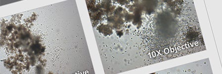
Is the scoring of CFU assays subjective?
Yes, the scoring of CFU assays can be subjective. Some of the most common and particularly challenging phenomena encountered when scoring CFU colonies is the presence of colonies with multiple foci or clusters, which can be scored as separate colonies and, thus, erroneously skew the total counts to higher CFU numbers. This, in turn, may lead to an overestimation of HSPC graft potency. Conversely, high plating concentrations can result in overlapping and underdeveloped colonies that can lead to under-scoring of CFUs. Training has been shown to improve consistency between manual counters with regard to both identification of colony number as well as lineage identification of each colony type.
I get motion sickness counting the colonies. How can I alleviate this problem?
Individuals new to counting CFU assays commonly experience motion sickness when counting colonies in a CFU assay. Ways to avoid this include:
Limiting the time spent at the microscope to just one hour at a time to start.
Counting in vertical rows by moving the stage control knob up and down rather than side-to-side across the dish.
Starting slow: start with no more than an hour of counting. With practice, you will be able to move on to counting colonies for a few hours at a time.
Taking breaks! Even once you’re feeling confident, it’s important to take a break every hour to stretch and look into the distance to relax your eyes.
If you have tips on preventing motion sickness when counting colonies that you’d like to share, let us know! Email us your tips at techsupport@stemcell.com.
How can I learn to count CFU numbers accurately and reproducibly?
The best way to learn to count CFU numbers accurately is with lots of practice! Here are some tips to help you get started:
At first, spend 1 - 2 hours per day, several days per week learning to recognize the different CFU types and to count accurately. Count the same cultures on different days. Cultures placed at 33°C, 5% CO2 and ≥ 95% humidity will maintain good morphology for a few more days in addition to the initial culture period.
Carry out comparative counting with qualified colleagues.
Ask us! Email pictures of your colonies to Product & Scientific Support at techsupport@stemcell.com to see that you’re culturing and scoring your colonies correctly.
Is it possible to characterize the subsets of the granulocytic lineage differentiated by CFU assay?
The granulocytic lineage consists of three mature cell types: neutrophils, basophils, and eosinophils. The only way to truly identify the frequency of these in peripheral blood is based on histological staining with hematoxylin and eosin (H&E) and evaluation of relevant morphological features that distinguishes these three types based on their size, color, and shape of the nucleus.
In a CFU assay, one only has the size of the mature cells to help identify the progenitor type and cannot visualize the shape of the nuclei. The shape of the nuclei is key in determining the difference between, for example, granulocytes and monocytes. The only way to identify the composition of the cells within a CFU-GM colony is to pluck the colony and evaluate the mature cells within the colony following a cytospin and subsequent H&E staining. However, most colonies are a heterogeneous mix of immature and mature cells that represents the lineage potential of the progenitor.
Alternatively, cells plucked from individual colonies can be stained with cell surface markers that are characteristic for the various mature cell types. However, this approach often involves staining of multiple surface markers to be definitive. Historically, histological staining, cell-surface expression by flow cytometry, and cytokine requirements have all been used to confirm lineage specific progenitor types.
Can I leave the human CFU cultures for longer than 14 days?
Human CFU cultures should not be left for much longer than 14 days. All human CFU cultures in standard MethoCult™ media, such as MethoCult™ H4435 Enriched, should be counted around days 13 -15.
The cells can't stay in culture much longer primarily because the nutrients in the media will be depleted after this time. All the cells will start to die, including the primitive progenitors you are interested in identifying.
In addition, we do not recommend analyzing CFU cultures after more than 15 days as colonies either:
Continue to grow, making it difficult to identify and enumerate accurately due to the resultantas growth results in overcrowding, merging, and overlapping, as is common with myeloid colonies, or
Begin to die and disintegrate, as is common with small erythroid colonies.
However, if you cannot count the CFU cultures within the recommended (13 -~15 day) time frame and you are using standard MethoCult™ media, please advise for them to transfer the CFU culture trays to a 33°C incubator with 5% CO2 (95% humidity) for an additional 1 - 2 days. The lower temperature will not prevent cell death or inhibit proliferation, but will assist in maintaining colony morphology, so that the colonies can still be enumerated correctly.
Can't find the answer you are looking for? Reach out to us and one of our hematopoietic specialists will get back to you.
Related Resources
On-Demand Mouse Hematopoietic CFU Assay Course
Learn the basics of mouse hematopoiesis, how to set up the CFU assay in your own lab, and confidently classify and count the resulting colonies. Progress at your own pace with virtual lectures and step-by-step lab videos in this free course.
Watch this video for step-by-step instructions on how to set up the hematopoietic colony forming unit (CFU) assay using methylcellulose-based MethoCult™ medium.
Thank you for your interest in this product.
Please provide us with your contact information and your local representative
will contact you with a customized quote. Where appropriate, they can also assist you with a(n):
Estimated delivery time for your area
Product sample or exclusive offer
In-lab demonstration
By submitting this form, you are providing your consent to STEMCELL Technologies Canada Inc. and its subsidiaries and affiliates (“STEMCELL”) to collect and use your information, and send you newsletters and emails in accordance with our privacy policy. Please contact us with any questions that you may have. You can unsubscribe or change your email preferences at any time.
Item added to your cart
Frequently Asked Questions on Performing the CFU Assay