Human Hematopoietic Stem and Progenitor Cell Markers and Phenotyping Panels
Technical tip from our dedicated team of Product and Scientific Support specialists
Every good experiment starts with the right cells. Cell isolation is great for fast and easy isolation of particular cell types, but sometimes an experiment requires a bit more specificity. Flow cytometry is a key tool that has revolutionized our ability to interrogate highly specific cell populations. Hematopoietic stem cells (HSCs) can only be definitively identified and characterized through in vivo experiments that measure the ability of HSCs to reconstitute the hematopoietic system following transplantation. Hematopoietic progenitor cells can be measured by in vivo transplantation in addition to in vitro culture assays that measure the ability of progenitors to proliferate and differentiate. Multi-parameter flow cytometry is a very important tool to phenotypically subdivide these hematopoietic stem and progenitor cell (HSPC) populations on the basis of cell surface marker expression and understand the diverse functions of different subsets of this heterogeneous group of cells.
HSPC Markers
The best known marker of human HSPCs is the cell surface glycoprotein CD34. This marker is very useful for identifying and isolating HSPCs from bone marrow and blood as it is highly expressed on most, if not all, human HSPCs, but absent on mature blood cells. Most CD34+ cells do not express so-called lineage (Lin) markers, expressed on mature blood cells, such as CD2, CD3, CD11b, CD11c, CD14, CD16, CD19, CD24, CD56, CD66b and CD235. The population of CD34+ cells are heterogeneous in their expression of other markers, such as CD38, CD45RA, CD90 and CD49f. These markers can be used in combination to distinguish HSPC subsets that differ in their hematopoietic-reconstitution potential, as summarized in Table 1.
Table 1. HSC and Progenitor Cell Subsets and Markers
HSPC Phenotyping Panel Design
Table 2 shows a recommended panel of fluorescently labeled antibodies for HSPC subset analysis. This panel has been designed such that the most dimly expressed cell surface marker, CD90, is detected using the brightest fluorochrome, PE. PE can be used with flow cytometers equipped with a 488 nm blue laser and a 640 nm red laser and four or more fluorescence detectors, which are common in most laboratories or flow cytometry core facilities. Optimized settings for data acquisition, gating strategy and subset identification are instrument-dependent and need to be determined empirically. Below is a staining panel for immunophenotyping HSPCs, including corresponding antibodies, which have been tested in-house to provide optimal resolution and flexibility when analyzed on commonly used flow cytometers.Table 2. Panel for HSPC Phenotyping
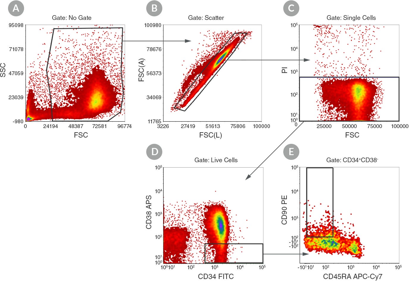
Figure 1. Gating Strategy for CD34+CD38-CD45RA-CD90+ HSC Subset

Explore our HSPC Research Resource Center to find more resources, including protocols, technical tips, webinars, wallcharts, and more.
Request Pricing
Thank you for your interest in this product. Please provide us with your contact information and your local representative will contact you with a customized quote. Where appropriate, they can also assist you with a(n):
Estimated delivery time for your area
Product sample or exclusive offer
In-lab demonstration


