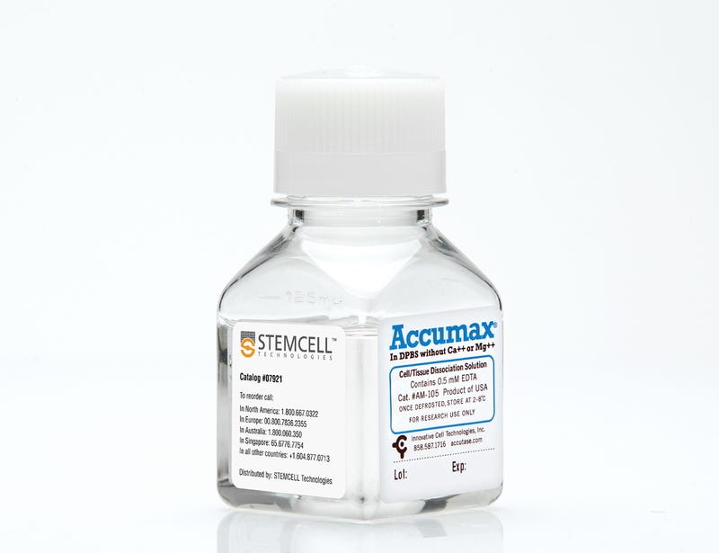
ACCUMAX™
Cell detachment solution
Request Pricing
Thank you for your interest in this product. Please provide us with your contact information and your local representative will contact you with a customized quote. Where appropriate, they can also assist you with a(n):
Estimated delivery time for your area
Product sample or exclusive offer
In-lab demonstration
By submitting this form, you are providing your consent to STEMCELL Technologies Canada Inc. and its subsidiaries and affiliates (“STEMCELL”) to collect and use your information, and send you newsletters and emails in accordance with our privacy policy. Please contact us with any questions that you may have. You can unsubscribe or change your email preferences at any time.
Overview
Protocols and Documentation
Find supporting information and directions for use in the Product Information Sheet or explore additional protocols below.
Resources and Publications
Publications (1)
Transcriptome Dynamics of Developing Photoreceptors in Three-Dimensional Retina Cultures Recapitulates Temporal Sequence of Human Cone and Rod Differentiation Revealing Cell Surface Markers and Gene Networks.
Stem cells (Dayton, Ohio) 2015 DEC
Abstract
The derivation of three-dimensional (3D) stratified neural retina from pluripotent stem cells has permitted investigations of human photoreceptors. We have generated a H9 human embryonic stem cell subclone that carries a green fluorescent protein (GFP) reporter under the control of the promoter of cone-rod homeobox (CRX), an established marker of postmitotic photoreceptor precursors. The CRXp-GFP reporter replicates endogenous CRX expression in vitro when the H9 subclone is induced to form self-organizing 3D retina-like tissue. At day 37, CRX+ photoreceptors appear in the basal or middle part of neural retina and migrate to apical side by day 67. Temporal and spatial patterns of retinal cell type markers recapitulate the predicted sequence of development. Cone gene expression is concomitant with CRX, whereas rod differentiation factor neural retina leucine zipper protein (NRL) is first observed at day 67. At day 90, robust expression of NRL and its target nuclear receptor NR2E3 is evident in many CRX+ cells, while minimal S-opsin and no rhodopsin or L/M-opsin is present. The transcriptome profile, by RNA-seq, of developing human photoreceptors is remarkably concordant with mRNA and immunohistochemistry data available for human fetal retina although many targets of CRX, including phototransduction genes, exhibit a significant delay in expression. We report on temporal changes in gene signatures, including expression of cell surface markers and transcription factors; these expression changes should assist in isolation of photoreceptors at distinct stages of differentiation and in delineating coexpression networks. Our studies establish the first global expression database of developing human photoreceptors, providing a reference map for functional studies in retinal cultures.
Related Products
Related Products

Legal Statement:
ACCUMAX is a trademark of Innovative Cell Technologies, Inc.
ACCUMAX is a trademark of Innovative Cell Technologies, Inc.
Quality Statement:
PRODUCTS ARE FOR RESEARCH USE ONLY AND NOT INTENDED FOR HUMAN OR ANIMAL DIAGNOSTIC OR THERAPEUTIC USES UNLESS OTHERWISE STATED. FOR ADDITIONAL INFORMATION ON QUALITY AT STEMCELL, REFER TO WWW.STEMCELL.COM/COMPLIANCE.
PRODUCTS ARE FOR RESEARCH USE ONLY AND NOT INTENDED FOR HUMAN OR ANIMAL DIAGNOSTIC OR THERAPEUTIC USES UNLESS OTHERWISE STATED. FOR ADDITIONAL INFORMATION ON QUALITY AT STEMCELL, REFER TO WWW.STEMCELL.COM/COMPLIANCE.
