Make more informed purchasing decisions with our new product availability and delivery estimate feature, now available on all product pages, in your cart, and during checkout.
Sign In
New to STEMCELL?
Register for an account to quickly and easily purchase products online and for one-click access to all educational content.
Thank you for your interest in this product.
Please provide us with your contact information and your local representative
will contact you with a customized quote. Where appropriate, they can also assist you with a(n):
Estimated delivery time for your area
Product sample or exclusive offer
In-lab demonstration
By submitting this form, you are providing your consent to STEMCELL Technologies Canada Inc. and its subsidiaries and affiliates (“STEMCELL”) to collect and use your information, and send you newsletters and emails in accordance with our privacy policy. Please contact us with any questions that you may have. You can unsubscribe or change your email preferences at any time.
Conveniently enrich the mitochondrial or cytosolic subcellular fractions from mammalian cells or tissues using the Mitochondrial Isolation Kit. This kit offers a quick mitochondrial isolation technique that has been verified for detection using western blot analysis and may be used to study apoptosis and signaling events between the two fractions.
Features:
• Fast and efficient enrichment of the active mitochondrial subcellular fraction
• Verified for use with western blot analysis
The Mitochondrial Isolation Kit offers two options to isolate the mitochondrial subcellular fraction. The first option is a reagent-based method for processing up to six samples concurrently. The second method utilizes Dounce homogenization, providing a two-fold increase in isolated mitochondria from a single sample. Both methods employ microcentrifugation to separate the mitochondrial fraction from the cytosolic fraction.
The enriched mitochondrial fractions preserve their biological activity and are compatible with downstream applications, including the study of mitochondrial respiration, apoptosis, inner mitochondrial membrane potential, and protein profiling (Itahana & Zhang Cancer Cell, 2008).
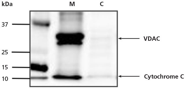
Figure 1. Western Blot of Mitochondrial Markers After Mitochondrial Isolation from Cultured Cells
Mitochondria were isolated from HEK293 cells with Mitochondrial Isolation Kit using Dounce homogenization. Mitochondrial (M) and cytosolic (C) fractions were analyzed via western blot for two mitochondrial proteins: voltage-dependent anion channels (VDAC) and cytochrome C. Results demonstrate that mitochondria remain intact after enrichment.
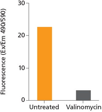
Figure 2. Mitochondria Isolated Using the Mitochondrial Isolation Kit Maintain Healthy Mitochondrial Function
Mitochondria were isolated from HEK293 cells using Mitochondrial Isolation Kit and stained with JC-1 (Iodide) dye (Catalog #100-0993) in the presence (grey bar) or absence (orange bar) of valinomycin. High levels of fluorescence indicate that healthy mitochondrial function is retained after isolation.
This product is designed for use in the following research area(s) as part
of the highlighted workflow stage(s). Explore these workflows to learn more about the other products we
offer to support each research area.