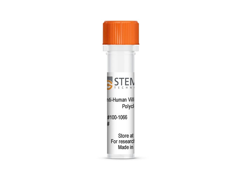
Anti-Human Villin 1 Antibody, Polyclonal

Rabbit polyclonal antibody against human villin 1

Anti-Human Villin 1 Antibody, Polyclonal

Rabbit polyclonal antibody against human villin 1

From: 425 USD
Catalog #
(Select a product)
Rabbit polyclonal antibody against human villin 1
Add to Wish List

Overview

This rabbit polyclonal antibody reacts with human villin 1, a calcium-regulated protein expressed in epithelial cells. Villin 1 is organized into six conserved domains, which make up the villin core, and a C-terminal headpiece domain, which contains an F-actin binding site. Use this polyclonal antibody in applications requiring detection of villin 1, such as western blotting (WB) assays and immunohistochemistry (IHC). It is verified for use with immunocytochemistry (ICC).

Villin 1 is an important player in the reorganization of microvillar actin filaments and is involved in actin nucleation, polymerization, and severing. It is a key component in the growth and development of intestinal epithelial cells, regulating cell morphology, growth, and apoptosis. Villin 1 has been suggested as a predictive factor in some adenocarcinomas of the endocervix, colon, and lungs. Abnormal villin expression has also been associated with biliary atresia and cholestasis.
Subtype
Primary Antibodies
Target Antigen
Villin1
Alternative Names
D2S1471, VIL1, VIL
Reactive Species
Human
Conjugation
Unconjugated
Host Species
Rabbit
Cell Type
Intestinal Cells
Species
Human
Application
Immunocytochemistry, Immunofluorescence, Immunohistochemistry, Western Blotting
Area of Interest
Organoids
Clone
Not applicable
Gene ID
7429
Isotype
Not applicable

Data Figures

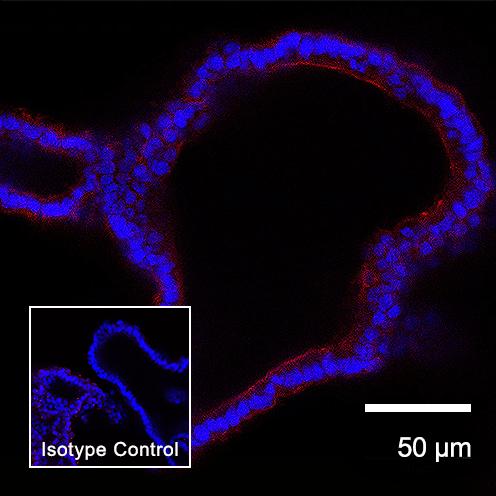
Figure 1. Anti-Human Villin 1 Antibody, Polyclonal is Suitable for Staining Human Intestinal Organoids

H9 human intestinal organoids were cultured using STEMdiff™ Intestinal Organoid Kit (Catalog # 05140), then fixed and labeled with Anti-Human Villin 1 Antibody, Polyclonal, followed by Goat Anti-Rabbit IgG (H+L) Antibody, Polyclonal, iFluor™ 568 (Catalog #100-1083). Nuclei were counter-stained with DAPI (blue). Inset shows cells labeled with a rabbit IgG isotype control antibody, followed by Goat Anti-Rabbit IgG (H+L) Antibody, Polyclonal, iFluor™ 568 (with DAPI staining).

Protocols and Documentation

Find supporting information and directions for use in the Product Information Sheet or explore additional protocols below.

Document Type
Product Name
Catalog #
Lot #
Language
Catalog #
100-1066
Lot #
All
Language
English
Document Type
Safety Data Sheet
Catalog #
100-1066
Lot #
All
Language
English

Applications

This product is designed for use in the following research area(s) as part of the highlighted workflow stage(s). Explore these workflows to learn more about the other products we offer to support each research area.