EasySep™ PE Positive Selection Kit II

Immunomagnetic positive selection of PE-conjugated antibody labeled cells
EasySep™ PE Positive Selection Kit II

Immunomagnetic positive selection of PE-conjugated antibody labeled cells

From: 605 USD
Catalog #
(Select a product)
Immunomagnetic positive selection of PE-conjugated antibody labeled cells
Add to Wish List

Product Advantages


  • Fast and easy-to-use

  • No columns required

What's Included

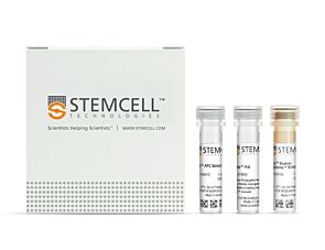
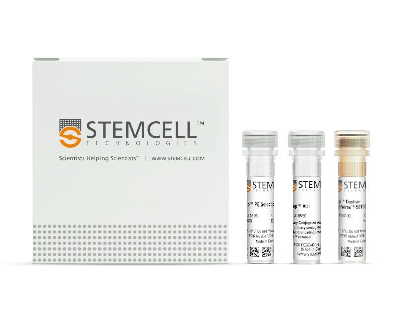
  • EasySep™ PE Positive Selection Kit II (Catalog #17684)
    • EasySep™ PE Selection Cocktail, 1 mL
    • EasySep™ Dextran RapidSpheres™, 1 mL
    • RoboSep™ Vial For Primary Conjugated Antibody (not required for manual use), 1 vial
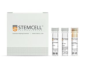
  • RoboSep™ PE Positive Selection Kit II (Catalog #17684RF)
    • EasySep™ PE Selection Cocktail, 1 mL
    • EasySep™ Dextran RapidSpheres™, 1 mL
    • RoboSep™ Vial For Primary Conjugated Antibody (not required for manual use), 1 vial
    • RoboSep™ Buffer (Catalog #20104)
    • RoboSep™ Filter Tips (Catalog #20125) x 2
Products for Your Protocol
To see all required products for your protocol, please consult the Protocols and Documentation.

Overview

Isolate highly purified phycoerythrin (PE)-conjugated antibody-labeled cells from any single-cell suspension samples by immunomagnetic positive selection, with the EasySep™ PE Positive Selection Kit II. Widely used in published research for more than 20 years, EasySep™ combines the specificity of monoclonal antibodies with the simplicity of a column-free magnetic system.

In this EasySep™ positive selection procedure, desired cells are labeled with antibody complexes recognizing PE and magnetic particles. Labeled cells are separated using an EasySep™ magnet and by simply pouring or pipetting off the unwanted cells. The cells of interest remain in the tube. Following magnetic cell isolation, the desired PE positive cells are ready for downstream applications.

This product replaces the EasySep™ PE Positive Selection Kit (Catalog #18557) for even faster cell isolations.

Learn more about how immunomagnetic EasySep™ technology works or how to fully automate immunomagnetic cell isolation with RoboSep™. Explore additional products optimized for your workflow, including culture media, supplements, antibodies, and more.

Magnet Compatibility
• EasySep™ Magnet (Catalog #18000)
• “The Big Easy” EasySep™ Magnet (Catalog #18001)
• EasyEights™ EasySep™ Magnet (Catalog #18103)
• RoboSep™-S (Catalog #21000)
Subtype
Cell Isolation Kits
Cell Type
Other
Species
Non-Human Primate, Other, Rat
Sample Source
Bone Marrow, Buffy Coat, Cord Blood, Leukapheresis, Other, PBMC, Spleen, Whole Blood
Selection Method
Positive
Application
Cell Isolation
Brand
EasySep, RoboSep
Area of Interest
Immunology

Protocols and Documentation

Find supporting information and directions for use in the Product Information Sheet or explore additional protocols below.

Document Type
Product Name
Catalog #
Lot #
Language
Catalog #
17684
Lot #
All
Language
English
Catalog #
17684RF
Lot #
All
Language
English
Document Type
Safety Data Sheet 1
Catalog #
17684
Lot #
All
Language
English
Document Type
Safety Data Sheet 2
Catalog #
17684
Lot #
All
Language
English
Document Type
Safety Data Sheet 1
Catalog #
17684RF
Lot #
All
Language
English
Document Type
Safety Data Sheet 2
Catalog #
17684RF
Lot #
All
Language
English
Document Type
Safety Data Sheet 3
Catalog #
17684RF
Lot #
All
Language
English

Resources and Publications

Publications (11)

Anti-endothelin receptor A autoantibodies associate with immunovascular risk and activate endothelial signalling in SLE. M. McCrorey et al. Lupus science & medicine 2026 Jul

Abstract

OBJECTIVE: Cardiovascular disease in SLE is not fully explained by traditional risk factors, supporting a role for disease-specific immunovascular mechanisms. Autoantibodies targeting endothelin receptor A (ETAR) and endothelin receptor B (ETBR) have been implicated in vascular injury in other autoimmune diseases, but their clinical and functional relevance in SLE remains unclear. We tested whether ET receptor autoantibodies associate with endothelial activation and directly stimulate ET receptor signalling in SLE. METHODS: Plasma anti-ETAR and anti-ETBR autoantibodies and soluble vascular cell adhesion molecule-1 (sVCAM-1) were measured in a pilot cohort of women with SLE and non-SLE controls and in an independent validation cohort stratified by SLE and hypertension (HTN) status. Multivariable models adjusted for demographic variables, blood pressure and medication use. Mechanistic studies were performed in primary human renal endothelial cells (HRECs) using live-cell calcium (Ca2+) imaging, ET receptor antagonists and receptor-directed blocking peptides. RESULTS: Anti-ETAR and anti-ETBR autoantibodies were elevated in SLE across both cohorts, with the highest level observed in SLE with HTN. Anti-ETAR demonstrated the strongest association with sVCAM-1 and independently identified women with the highest endothelial activation burden beyond clinical covariates and anti-double-stranded DNA. In primary HRECs, SLE-derived IgG induced Ca2+ flux that was attenuated by ET receptor antagonism. Blockade of the ETAR extracellular loop 2 domain reduced IgG-mediated Ca2+signalling, supporting a non-canonical mechanism of endothelial ETAR activation. CONCLUSIONS: Anti-ETAR autoantibodies associate with immunovascular risk and endothelial activation in SLE and directly stimulate endothelial signalling through a non-canonical ETAR-dependent mechanism. These findings support the use of anti-ETAR autoantibodies as clinically relevant biomarkers and potential novel contributors to vascular dysfunction in SLE.
High Treg and PMN-MDSC densities are a hallmark of tertiary lymphoid structures in fatal cases of cervical cancer L. A. Syding et al. Journal for Immunotherapy of Cancer 2025 Sep

Abstract

BackgroundHigh densities of tertiary lymphoid structures (TLSs) are associated with improved clinical outcomes in various malignancies, including human papillomavirus (HPV)-associated head and neck squamous cell carcinoma (HNSCC). However, the role of TLSs in shaping antitumor immunity in HPV-induced cervical cancer (CESC) remains unclear. Therefore, we analyzed the density, composition, and prognostic impact of TLSs in patients with CESC as well as patients with HNSCC.MethodsMultiplex immunofluorescence, immunohistochemistry, and spatial transcriptomics were used to analyze TLS density and composition in HNSCC and CESC tissue sections with respect to patient prognosis. The spatial approach was supplemented by flow cytometry-based analysis of the polymorphonuclear myeloid-derived suppressor cell (PMN-MDSC) phenotype in freshly resected primary tumor tissues.ResultsAlthough both indications were associated with HPV infection, we confirmed a positive correlation between TLS density and improved overall survival only in patients with HNSCC. The TLS composition differed markedly between HNSCC and CESC samples, with a shift toward high regulatory T cell (Treg) and PMN-MDSC abundance in CESC samples. The highest Treg and PMN-MDSC levels were observed in patients with CESC who died of the disease. CESC-infiltrating PMN-MDSCs showed high arginase 1 expression, which correlated with diminished T-cell receptor (TCR)ζ chain expression in CESC-infiltrating T cells. Additionally, the high number of PMN-MDSCs in TLSs was associated with the absence of HPV-specific T cells in CESC.ConclusionsUnlike in HNSCC, the composition of TLSs, rather than their quantity, was associated with the overall survival of patients with CESC. High numbers of Tregs and PMN-MDSCs infiltrating immature TLSs prevail in patients with CESC who succumbed to the disease and seem to affect tumor-specific immune responses.
CD56 on intratumoral NK cells: orchestrating NK cell-mediated anti-tumor effects in bladder cancer Z. Hassouneh et al. Neoplasia (New York, N.Y.) 2025 May

Abstract

Highlights•CD56bright NK cells correlate with better survival outcomes in bladder cancer.•Deletion of CD56 impairs NK cell cytotoxicity against bladder cancer cells.•CD56 deletion affects NK cell migration and adhesion to tumor cells.•CD56 expression on BCa cells may predict efficacy of NK cell-based immunotherapy. Bladder cancer (BCa) exhibits favorable responses to immunotherapy, but a significant percentage of patients fail to show a response owing to an inadequate tumor-immune landscape. We previously showed that NK cells are one of the predominant tumor-infiltrating lymphocytes in BCa and correlate with improved patient survival. However, that link was observed only with CD56bright NK cells while the CD56dim subset exhibited reduced cytotoxicity and higher accumulation in advanced BCa stages. The role of CD56 in NK cell functionality in BCa, however, remains unclear. Using flow cytometry and cytotoxicity assays, we demonstrated a significant decrease in cytotoxicity and activation of NK92 cells against BCa upon CD56 deletion. Further, migration assays and atomic force microscopy showed CD56 deletion impaired NK92 cell migration and adhesion to bladder tumor cells, reducing NK92 cell-mediated apoptosis of BCa cells. Prolonged exposure to bladder tumors led to CD56 loss in NK92 cells, suggesting tumor-induced NK92 cell dysfunction via CD56 reduction, consistent with our previous findings. Confocal microscopy revealed an overlap of CD56 and phosphorylated Pyk2, a critical kinase at the tumor-immune synapse, potentially mediating the downstream cytotoxicity effects. Blocking Pyk2 phosphorylation decreased CD56-mediated NK92 cell activation and reduced NK92 cell-mediated cytotoxicity against BCa. Finally, we showed that CD56 is also expressed by BCa cells and may be a predictive biomarker for NK cell-based immunotherapy, with its shedding indicating a mechanism for NK cell evasion. Our study identifies a novel innate-immune axis in BCa, leading to a better understanding of intratumoral NK cell biology and advancing NK cell-targeted treatments. Graphical abstractImage, graphical abstract