Generation of Megakaryocytes from Pluripotent Stem Cells Using STEMdiff™ Megakaryocyte Kit
- Document # 27226
- Version 1.0.1
- Oct 2023
Background
Culture methods to differentiate human embryonic stem (ES) cells and induced pluripotent stem (iPS) cells to megakaryocytes and platelets are important for studying megakaryopoiesis and platelet production and the diseases that affect them. Platelet transfusion is an effective treatment for thrombocytopenia-related diseases, yet a shortage of supply and limited shelf-life remain challenging. Having a renewable source of non-donor-derived platelets could alleviate the strain on precious donor samples.
Existing methods for generating megakaryocytes from hPSCs are technically challenging, involve multiple culture steps, have low cell yields, and lack reproducibility between cell lines and experiments. Here, we describe a simple two-step, serum- and feeder-free culture system to robustly generate polyploid megakaryocytes with high yield and purity, across multiple hPSC lines, using the STEMdiff™ Megakaryocyte Kit. These polyploid megakaryocytes are also capable of shedding platelet-like particles (PLPs). By providing a safe and reliable supply of megakaryocytes, STEMdiff™ Megakaryocyte Kit is well-positioned for megakaryopoiesis and thrombopoiesis research, gene-editing applications, and cellular therapies.
Differentiate Human ES and iPS Cells to Polyploid Megakaryocytes
STEMdiff™ Megakaryocyte Kit is designed for the serum-free and feeder-free differentiation of human embryonic stem (ES) and induced pluripotent stem (iPS) cells to polyploid megakaryocytes capable of shedding PLPs and expressing CD41a and CD42b in a simple two-dimensional, 17-day, two-stage protocol, as shown in Figure 1. The kit comprises two culture media—STEMdiff™ Hematopoietic Basal Medium and StemSpan™ SFEM II—and the following three supplements:
- STEMdiff™ Hematopoietic Supplement A: Promotes mesodermal specification of hPSCs
- STEMdiff™ Megakaryocyte Supplement MK1: Promotes differentiation to and expansion of megakaryocyte-biased hematopoietic progenitors
- STEMdiff™ Megakaryocyte Supplement MK2: Promotes differentiation of hematopoietic progenitor cells to mature megakaryocytes
Why Use STEMdiff™ for Generating Megakaryocytes?
- Reduce variability with a serum- and feeder-free formulation
- Achieve robust generation of megakaryocytes across multiple ES and iPS cell lines
- Generate high yields of megakaryocytes per input hPSC
- Amenable to gene editing and to large-scale culture (feeder-free)
| Product Name | Size | Catalog # |
|---|---|---|
| STEMdiff™ Megakaryocyte Kit | 1 Kit | 100-0900 |
| Kit Components | Size | Catalog # |
| STEMdiff™ Hematopoietic Basal Medium | 120 mL | 05311 |
| STEMdiff™ Hematopoietic Supplement A (200X) | 225 μL | 05312 |
| STEMdiff™ Megakaryocyte Supplement MK 1 (10X) | 10 mL | 100-0901 |
| STEMdiff™ Megakaryocyte Supplement MK 2 (10X) | 10 mL | 100-0902 |
| StemSpan™ SFEM II | 100 mL | 09605 |
Protocol for Generating Megakaryocytes
The following protocol is recommended for the expansion and megakaryocytic differentiation of hPSCs maintained in mTeSR™1, mTeSR™ Plus, or TeSR™-AOF™.
Using this protocol, cell yields may reach between 223 - 425 CD41a⁺CD42b⁺ megakaryocytes per input hPSC, with purity ranging from 56 - 77% CD41a⁺CD42b⁺ cells. The yields and frequencies may vary between experiments depending on the cell line and condition of the cells at the start of the differentiation culture. Depending on your specific objectives, further protocol optimization (e.g. testing of different plating densities, culture periods, and feeding and replating schedules) may be required.

Figure 1. Megakaryocyte Differentiation Protocol
The 17-day protocol includes two main stages: a 12-day stage to differentiate human embryonic stem (hES) or induced pluripotent stem (hiPS) cells into megakaryocyte-biased hematopoietic progenitor cells (HPCs), and a 5-day stage to further differentiate hES or hiPS cell-derived HPCs into mature megakaryocytes (MKs). On Day -1, hES or hiPS cells are plated as aggregates (100 - 200 μm diameter, ~100 cells per aggregate) at a density of 10 ‑ 20 aggregates/cm2 in mTeSR™1, mTeSR™ Plus, or TeSR™-AOF on Corning® Matrigel®-coated plates. After attaching overnight and confirming the number of adhered colonies is within 4 - 10 colonies/cm2, mesoderm induction is initiated by replacing TeSR™ medium with Medium A (STEMdiff™ Hematopoietic Basal Medium + STEMdiff™ Hematopoietic Supplement A). On Day 3, the medium is changed to Medium MK1 (STEMdiff™ Hematopoietic Basal Medium + STEMdiff™ Megakaryocyte Supplement MK1) for endothelial-to-hematopoietic transition (EHT) and hematopoietic specification. During this phase, hES or hiPS cell-derived HPCs emerge from an adherent layer of endothelial cells and are released into suspension. On Day 12, HPCs in suspension are harvested and replated in Medium MK2 (StemSpan™ SFEM II + STEMdiff™ Megakaryocyte Supplement MK2) at a density of 1 - 3.5 x 10⁵ cells/mL and cultured for 5 days to generate mature MKs.
This procedure has been optimized for use with human ES and iPS cells; refer to the Product Information Sheet (PIS; Document #10000013343) for complete instructions.
1. Passage hPSC aggregates and set up for differentiation as per the protocol in the PIS.
Note: For complete instructions on maintaining high-quality human ES and iPS cells and for coating plates with Corning® Matrigel®, refer to the Technical Manual for mTeSR™1, mTeSR™ Plus, or TeSR™ E8™, available at www.stemcell.com, or contact us to request a copy.
Hematopoietic Differentiation Protocol
Note: Throughout the protocol, warm all media to room temperature (15 - 25°C) before use. Do not leave the media at room temperature for extended periods of time.
- On Day 0, confirm that 16 - 40 colonies/well are adhered to the cultureware (4 - 10 colonies/cm²; this protocol is based on 12-well plates, but can be adapted for other cultureware sizes). Aspirate medium from wells.
- Add 1 mL of Medium A (STEMdiff™ Hematopoietic Basal Medium + STEMdiff™ Hematopoietic Supplement A) per well. Store remaining Medium A at 2 - 8°C until required.
- Incubate at 37°C and 5% CO2 for 2 days.
- On Day 2, gently remove 0.5 mL of medium from each well and discard. Gently add 0.5 mL of Medium A per well.
- Incubate at 37°C for 24 hours.
- On Day 3, aspirate medium from wells. Gently add 1 mL of Medium MK1 (STEMdiff™ Hematopoietic Basal Medium + STEMdiff™ Megakaryocyte Supplement MK1) per well. Store remaining Medium MK1 at 2 - 8°C until required.
- Incubate at 37°C for 2 days.
- Perform a full-medium change with Medium MK1 on Days 5, 7, and 10, being careful not to disturb the floating cell population, as per the PIS.
- Incubate at 37°C for 2 days after each medium change.
- On day 12, harvest hematopoietic cells as follows:
- Pipette the cells up and down to wash the well and remove the suspension cells from the adherent cell layer.
- Add 1 mL of StemSpan™ SFEM II to the well. Triturate vigorously in the well and add to the collection tube.
- Repeat steps 10a and 10b for each well.
- Centrifuge collection tubes at 300 x g for 5 minutes at room temperature (15 - 25°C).
- Remove and discard the supernatant.
- Resuspend the cell pellet in StemSpan™ SFEM II and perform a viable cell count using Trypan Blue and a hemocytometer.
Megakaryocytic Differentiation Protocol
- On Day 12, add 1 mL of Medium MK2 (StemSpan™ SFEM II + STEMdiff™ Megakaryocyte Supplement MK2) to each well of a tissue culture-treated 12-well plate.
- Add harvested cells (from step 10 of the Hematopoietic Differentiation Protocol) to each well at 100,000 - 350,000 cells/mL.
- Incubate at 37°C for 5 days.
- On day 17, harvest megakaryocytes as follows:
- Pipette the cells up and down to wash the wells.
- Transfer the cell suspension to a collection tube.
- Centrifuge collection tubes at 300 x g for 5 minutes at room temperature (15 - 25°C).
- Remove and discard the supernatant.
- Resuspend the cell pellet in desired medium and perform a viable cell count for analysis or downstream assay. Purity of CD41a+CD42b+ megakaryocytic cell population can be determined by flow cytometry.
Results

Figure 2. Megakaryocyte Differentiation Protocol
hES and hiPS cells were induced to differentiate to megakaryocyte-erythroid biased HPCs following the protocol described in Figure 1. On Day 12, cells were harvested from the supernatant and analyzed for expression of CD41a, CD42b, CD34, and GlyA by flow cytometry. Dead cells were excluded by light scatter profile and propidium iodide (PI) staining. (A) Representative flow cytometry plots for hES-derived (H9) cells on Day 12. The cells show high levels of CD41a and CD42b as well as CD34 and GlyA expression, indicating that the protocol supports differentiation of hPSCs to megakaryocyte-erythroid progenitors by Day 12. (B) Frequencies and numbers of CD41a⁺CD42b⁺ cells per input cell for two hES cell lines (H1 and H9) and two hiPS cell lines (WLS-1C and STiPS-R038). The average frequency of viable CD41a⁺CD42b⁺ cells on Day 12 ranged between 33% and 62%. The average yield of CD41a⁺CD42b⁺ cells generated per input cell ranged between 72 and 174. Data are shown as mean ± SEM (n = 7 for H1, n = 20 for H9, n = 19 for WLS-1C, n = 7 for STiPS-R038).
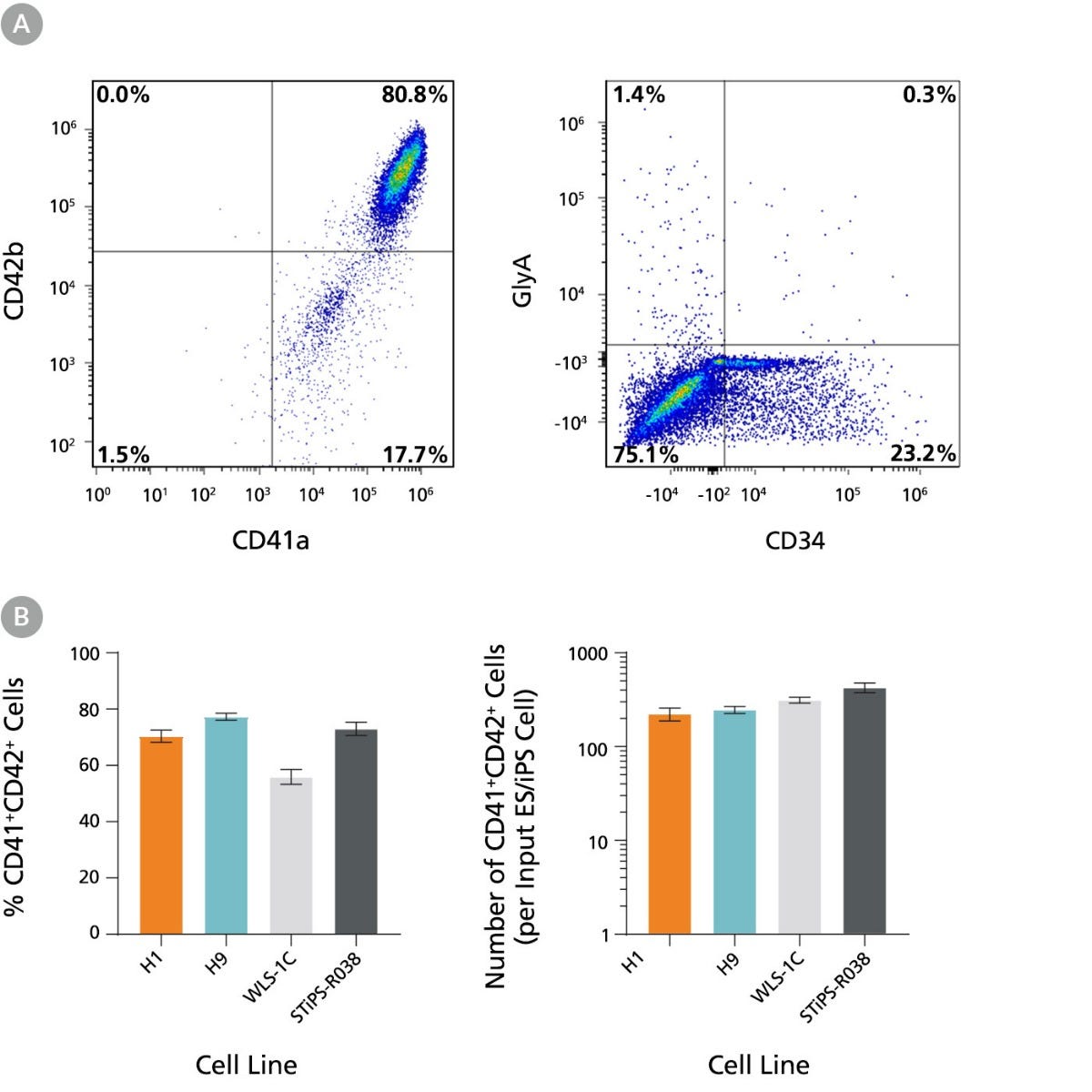
Figure 3. hPSC-Derived HPCs Efficiently Expand and Differentiate to CD41a+CD42b+ Megakaryocytes During an Additional 5 Days of Culture
hPSC-derived HPCs on Day 12 were cultured for 5 additional days in Medium MK2 to promote differentiation into mature MKs following the protocol described in Figure 1. Cells were harvested and analyzed for expression of CD41a, CD42b, CD34, and GlyA by flow cytometry. Dead cells were excluded by light scatter profile and PI staining. (A) Representative flow cytometry plots for hES-derived (H9) cells on Day 17. The cells showed high levels of CD41a and CD42b and low levels of GlyA and CD34 markers, indicating differentiation to megakaryocytes. (B) Frequencies and numbers of CD41a⁺CD42b⁺ MKs per input cell for two hES cell lines (H1 and H9) and two hiPS cell lines (WLS-1C and STiPS-R038). The average frequency of viable CD41a⁺CD42b⁺ cells on day 17 ranged between 56% and 77%. The average yield of CD41a⁺CD42b⁺ MKs generated per input cell ranged between 223 and 425. Data are shown as mean ± SEM (n = 12 for H1, n = 29 for H9, n = 27 for WLS-1C, n = 12 for STiPS-R038).

Figure 4. hPSC-Derived Megakaryocytes Generated Using STEMdiff™ Megakaryocyte Kit Are Polyploid
hPSC-derived MKs obtained using STEMdiff™ Megakaryocyte Kit display mature and adult-like features: cellular enlargement and polyploidization. (A) A representative bright-field image taken on Day 17 showing large MKs derived from H1 cells (10X magnification). (B) Representative immunofluorescence images taken on Day 17 showing that CD41a⁺ MKs derived from H1 and WLS-1C cells are polyploid (20X and 63X magnification, respectively). The cells were formaldehyde-fixed and stained with a fluorescein-conjugated antibody against surface marker CD41a (green), and DAPI (blue). (C) A representative cytospin of MKs derived from H9 cells on Day 17 showing high ploidy (40X magnification, May-Grunwald Giemsa stain). (D) Representative flow cytometry histogram and scatter plot showing the DNA ploidy profile of ethanol-fixed MKs derived from H9 cells on Day 17. The DNA content was determined by the quantity of PI staining, with different peaks on the histogram representing 2N, 4N, and 8N+ cells. Ploidy analysis was done on gated CD41a⁺ cells. (E) Ploidy distribution of MKs generated from two hES cell lines (H1 and H9) and two hiPS cell lines (WLS-1C and STiPS-R038). The average ploidy distributions of CD41a+ cells on Day 17 were 66%, 24.5%, and 9.5% for 2N, 4N, and 8N+, respectively. Data are shown as mean ± SEM (n = 6 for H1, n = 28 for H9, n = 19 for WLS-1C, n = 10 for STiPS-R038).

Figure 5. hPSC-Derived Megakaryocytes Generated Using STEMdiff™ Megakaryocyte Kit Yield Platelet-Like Particles
hPSC-derived MKs obtained using STEMdiff™ Megakaryocyte Kit are capable of proplatelet formation to yield functional platelet-like particles (PLPs). (A) A representative bright-field image taken on Day 17 showing MKs derived from H1 cells formed proplatelets (long cytoplasmic protrusions;10X magnification). (B) Representative flow cytometry forward/side scatter profile of MKs and PLPs and histogram of PLPs derived from H9 cells on Day 17. The PLP gate is based on control platelets (PLTs) prepared from fresh blood. Cells were also stained with antibodies against CD41a, CD45, and GlyA for PLP characterization and enumeration. PLPs showed a high level of CD41a expression (and no CD45 and GlyA expression; data not shown) as in control PLTs. Grey filled histogram represents CD41a Fluorescence Minus One (FMO) control. (C) Numbers of PLPs generated per MK on Day 17 for two hES cell lines (H1 and H9) and two hiPS cell lines (WLS-1C and STiPS-R038). PLPs and MKs were enumerated based on the number of CD41a⁺CD45⁻GlyA⁻ cells in the PLP gate and viable CD41a⁺ cells in the MK gate, respectively. The average yield of PLPs generated per MK ranged between 3.2 and 5.1. Data are shown as mean ± SEM (n = 12 for H1, n = 28 for H9, n = 27 for WLS-1C, n = 12 for STiPS-R038).

Figure 6. STEMdiff™ Megakaryocyte Kit Produces More Megakaryocytes and Platelet-Like Particles than Other Published Protocols
hES and hiPS cells were differentiated into MKs using STEMdiff™ Megakaryocyte Kit and using four different published protocols from the literature, with modifications. (A) Frequencies and numbers of CD41a⁺CD42b⁺ MKs per input cell for two hES cell lines (H1 and H9) and two hiPS cell lines (WLS-1C and STiPS-R038) were analyzed by flow cytometry, as shown in Figure 2. (B) Numbers of PLPs generated per input cell were enumerated as described in Figure 5. Compared to the published protocols, STEMdiff™ Megakaryocyte Kit produced 10- to 40-fold more CD41a⁺CD42b⁺ MKs and 6- to 23-fold more PLPs per input cell. Pp values were calculated using a one-way ANOVA followed by Dunnett’s post hoc test (*p < 0.05, **p < 0.01). Data are shown as mean ± SEM (n = 7 - 8).
Product Information
Materials Required but Not Included
| Product Name | Catalog # |
|---|---|
| Hausser Scientific™ Bright-Line Hemocytometer | 100-1181 |
| mTeSR™1 OR mTeSR™ Plus OR TeSR™-AOF | 85850 OR 100-0276 OR 100-0401 |
| Corning® Matrigel® hESC-Qualified Matrix | Corning 354277 |
| Gentle Cell Dissociation Reagent OR ReLeSR™ OR Dispase (1 U/mL) | 100-0485 OR 100-0484 OR 07923 |
| DMEM/F-12 with 15 mM HEPES | 36254 |
| Trypan Blue | 07050 |
| 12-well tissue culture-treated plates | e.g. 38052 |
| 96-well flat-bottom plate | e.g. 38022 |
Recommended Antibodies for Analysis
| Product Name | Catalog # |
|---|---|
| Anti-Human CD41a Antibody, Clone HIP8 | 60114 |
| Anti-human CD42b Antibody, Clone HIP1 | BioLegend 303912 |
Request Pricing
Thank you for your interest in this product. Please provide us with your contact information and your local representative will contact you with a customized quote. Where appropriate, they can also assist you with a(n):
Estimated delivery time for your area
Product sample or exclusive offer
In-lab demonstration


