
Ascorbic Acid
Antioxidant; Reducing agent
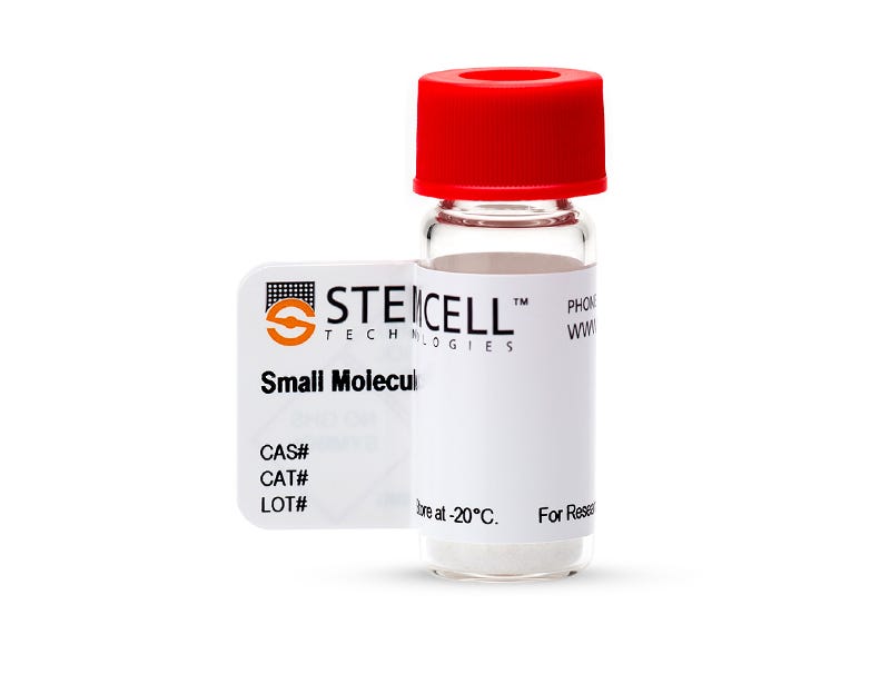
Request Pricing
Thank you for your interest in this product. Please provide us with your contact information and your local representative will contact you with a customized quote. Where appropriate, they can also assist you with a(n):
Estimated delivery time for your area
Product sample or exclusive offer
In-lab demonstration
By submitting this form, you are providing your consent to STEMCELL Technologies Canada Inc. and its subsidiaries and affiliates (“STEMCELL”) to collect and use your information, and send you newsletters and emails in accordance with our privacy policy. Please contact us with any questions that you may have. You can unsubscribe or change your email preferences at any time.
This site is protected by reCAPTCHA and the Google Privacy Policy and Terms of Service apply.
This site is protected by reCAPTCHA and the Google Privacy Policy and Terms of Service apply.
Overview
Protocols and Documentation
Find supporting information and directions for use in the Product Information Sheet or explore additional protocols below.
Resources and Publications
Publications (29)
Neural Cell Models of Specific Reading Disabilities: Identification of Cellular and Transcriptome Alterations.
Genes, brain, and behavior 2026 Oct
Abstract
Based on post-mortem studies, a long-standing hypothesis on the biological bases of specific reading disabilities (RD) has been that alterations in neuronal migration alter language neural circuits. However, there are questions concerning the original studies (small sample size, comorbidities). Further, this idea has not been strongly supported by current genetic studies. In this study, we test altered neural cell phenotypes using cells derived from two pairs of siblings strongly discordant for reading-probands with severe RD and their strong-reader siblings. We observed significant differences in neurosphere outgrowth between the neural precursor cells (NPCs) from the probands compared to their strong-reader siblings. Transcriptome analyses of adherent NPCs and neurons derived from the NPCs identified genes differentially expressed between the probands and siblings, including genes previously identified in neurodevelopmental disorders. Of note was the overexpression of the hormone IGF1 and the transcription factor OTX2, responsible for the opening and closing of critical windows in development and predicted to be involved in critical windows for language acquisition. Our results support altered transcriptome and cellular phenotypes in RD neural cells and suggest possible molecular mechanisms underlying the changes.
CDK2/9 blockade remodels chromatin and drives phenotypic plasticity in glioblastoma organoids.
Cell communication and signaling : CCS 2026 Oct
Abstract
BACKGROUND: Glioblastoma (GBM) remains a highly lethal brain tumor with limited therapeutic options and pervasive resistance to standard treatments. Cyclin-dependent kinases (CDKs), key regulators of cell cycle progression and transcription, are frequently dysregulated in GBM and contribute to tumor pathogenesis. METHODS: We employed a range of preclinical GBM models-including 2D cultures, 3D spheroids, patient-derived organoids (PDOs), and multicellular 3D bioprints-to evaluate the effects of the CDK2/9 inhibitor fadraciclib in comparison to the standard-of-care temozolomide. Endpoints included cell viability, stress responses, immune-modulatory effects, and transcriptomic reprogramming. RESULTS: Fadraciclib robustly reduces cell viability and invasion in all patient-specific 2D- and 3D models. This effect was independent of O6-methylguanine-DNA methyltransferase (MGMT) promoter methylation status, demonstrating superior efficacy to temozolomide. Mechanistically, fadraciclib disrupted lysosomal, mitochondrial, and endoplasmic reticulum integrity and increased γH2AX levels, indicating enhanced DNA double-strand breaks. Seahorse analysis demonstrated impaired mitochondrial bioenergetics following fadraciclib treatment. In a triple-culture blood-brain barrier (BBB) model, fadraciclib demonstrated effective BBB penetration and antitumoral activity against GBM cells, while exhibiting minor cytotoxicity toward brain-specific normal cells. In semi-autologous co-culture systems, fadraciclib inhibited GBM invasion, promoted intercellular communication via Connexin 43 upregulation in microglia, and activated peripheral T cells. Transcriptomic analyses of treated patient-derived organoids revealed widespread transcriptional reprogramming, including downregulation of oncogenic long non-coding RNAs (MALAT1, POM121L9), suppression of immune-related pathways, and upregulation of the tumor suppressor LZTS1. Increased histone-associated gene expression suggests chromatin remodeling and induction of a quiescent tumor cell state. Subtype analysis indicated a shift toward a proneural molecular signature. Notably, the antitumoral effects of fadraciclib were preserved in multicellular biomimetic bioprints, whereas nonmalignant astrocytes and microglia were not affected. Therapeutic activity also coincided with altered secretion profiles of extracellular vesicles (EV). EVs from reactive astrocytes contained higher amounts of Fas, which may act as modulator of neuroinflammation. CONCLUSION: CDK2/9 inhibition shows promise as a therapeutic strategy for GBM. Fadraciclib exhibits multifaceted anti-tumor activity and spares nonmalignant cells of the central nervous system, supporting its further development in therapies that target transcriptional and immune pathways, as well as quiescent tumor cell populations.
Consequences of the Novel ALS-Associated KIF5A Variant c.2993-6C > A for Exon 27 Splicing and Axonal Transport of SFPQ
Neurology: Genetics 2026 Mar
Abstract
Background and Objectives: Recent studies have identified variants in the kinesin family member 5A (KIF5A) gene that predispose to amyotrophic lateral sclerosis (ALS). These ALS-linked KIF5A variants lead to the exclusion of exon 27, resulting in the production of a mutated protein with an altered C-terminal region (KIF5A ΔExon27). Through whole genome sequencing, we identified a novel KIF5A intronic variant, rs1057522322 (c.2993-6C > A; chr12:57582596C > A, GRCh38.p14), in a family segregating ALS. Our goal is to investigate the effect of this variant on exon 27 splicing and to assess its functional consequences on KIF5A-mediated cargo transport. Methods: Induced pluripotent stem cells (iPSCs) were generated from siblings with and without the c.2993-6C > A variant. RT-PCR was performed on RNA extracted from iPSC-derived neurons to assess exon 27 splicing. Functional studies were conducted on iPSC-derived motor neurons (MNs). Results: RT-PCR confirmed that the c.2993-6C > A variant induced exon 27 skipping in KIF5A. Immunofluorescent staining showed that KIF5A ΔExon27 abolished the axonal interaction with splicing factor proline- and glutamine-rich, a cargo specifically transported by KIF5A. Under stress conditions, MNs carrying the c.2993-6C > A variant exhibited TDP-43 proteinopathy. Discussion: KIF5A intronic variant c.2993-6C > A could be a risk factor for ALS. KIF5A ΔExon27 impairs KIF5A-mediated cargo transport and contributes to ALS pathogenesis in a TDP-43–dependent manner.

Quality Statement:
PRODUCTS ARE FOR RESEARCH USE ONLY AND NOT INTENDED FOR HUMAN OR ANIMAL DIAGNOSTIC OR THERAPEUTIC USES UNLESS OTHERWISE STATED.
PRODUCTS ARE FOR RESEARCH USE ONLY AND NOT INTENDED FOR HUMAN OR ANIMAL DIAGNOSTIC OR THERAPEUTIC USES UNLESS OTHERWISE STATED.
