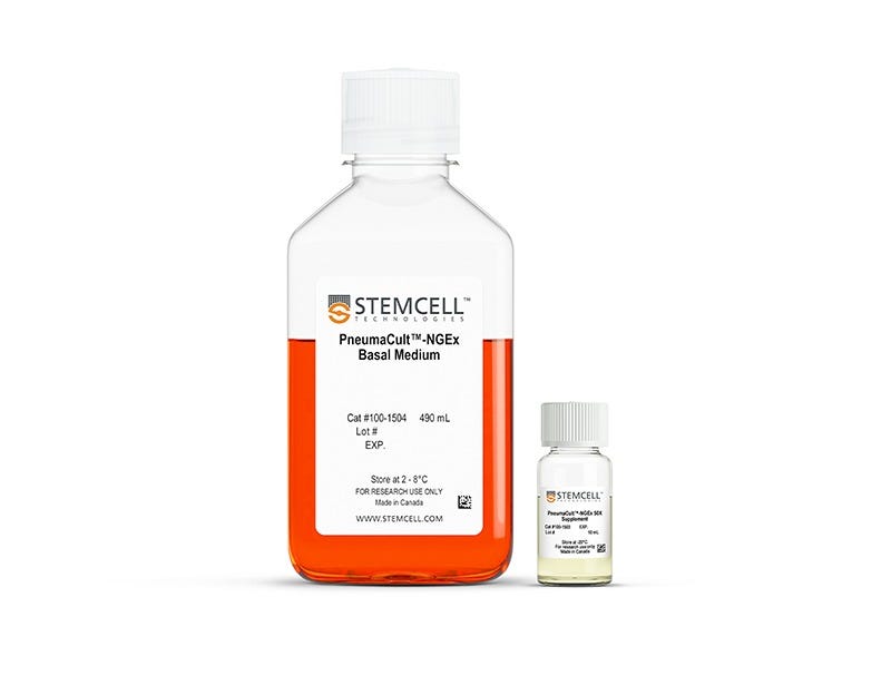
PneumaCult™-NGEx Medium
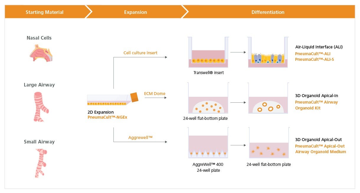
Serum- and BPE-free culture medium for the expansion of human large airway, small airway, and nasal epithelial cells
Request Pricing
Thank you for your interest in this product. Please provide us with your contact information and your local representative will contact you with a customized quote. Where appropriate, they can also assist you with a(n):
Estimated delivery time for your area
Product sample or exclusive offer
In-lab demonstration
-
 Animal Component-Free Cell Dissociation Kit
Dissociation kit for human stem and progenitor cells
-
 Hydrocortisone Stock Solution
Cell culture supplement
-
 D-PBS (Without Ca++ and Mg++)
Dulbecco’s phosphate-buffered saline without calcium and magnesium
What Our Scientist Says
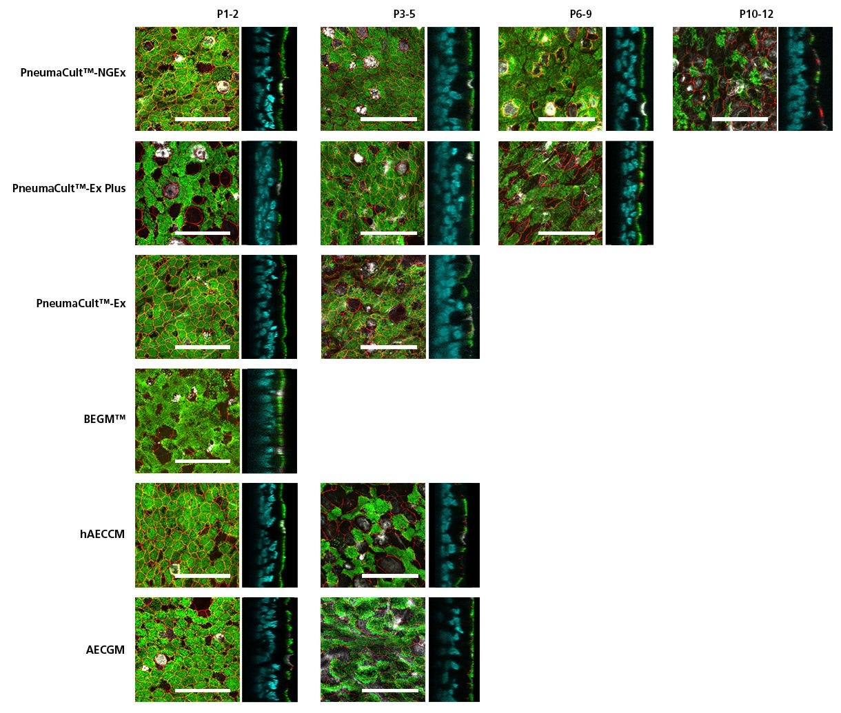
The development of our third-generation PneumaCult™ expansion medium reflects our commitment to continuous innovation and improvement. PneumaCult™-NGEx was designed to be the new Gold Standard in airway epithelial cell expansion and enable pulmonary scientists to push the boundaries of their work.

Overview
Data Figures


Protocols and Documentation
Find supporting information and directions for use in the Product Information Sheet or explore additional protocols below.
Applications
This product is designed for use in the following research area(s) as part of the highlighted workflow stage(s). Explore these workflows to learn more about the other products we offer to support each research area.
Resources and Publications
Educational Materials (9)


Related Products

PRODUCTS ARE FOR RESEARCH USE ONLY AND NOT INTENDED FOR HUMAN OR ANIMAL DIAGNOSTIC OR THERAPEUTIC USES UNLESS OTHERWISE STATED. FOR ADDITIONAL INFORMATION ON QUALITY AT STEMCELL, REFER TO WWW.STEMCELL.COM/COMPLIANCE.
