RosetteSep™ Human T Cell Enrichment Cocktail

Immunodensity negative selection cocktail

New look, same high quality and support! You may notice that your instrument or reagent packaging looks slightly different from images displayed on the website, or from previous orders. We are updating our look but rest assured, the products themselves and how you should use them have not changed. Learn more

RosetteSep™ Human T Cell Enrichment Cocktail

Immunodensity negative selection cocktail

From: 239 USD
Catalog #
(Select a product)
Immunodensity negative selection cocktail
Add to Wish List

Product Advantages


  • Fast and easy-to-use

  • Requires no special equipment or training

  • Isolated cells are untouched

  • Can be combined with SepMate™ for consistent, high-throughput sample processing

What's Included

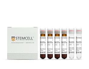
  • RosetteSep™ Human T Cell Enrichment Cocktail (Catalog #15021)
    • RosetteSep™ Human T Cell Enrichment Cocktail, 2 mL
  • RosetteSep™ Human T Cell Enrichment Cocktail (Catalog #15061)
    • RosetteSep™ Human T Cell Enrichment Cocktail, 5 x 2 mL
Products for Your Protocol

Overview

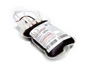
The RosetteSep™ Human T Cell Enrichment Cocktail is designed to isolate T cells from whole blood by negative selection. Unwanted cells are targeted for removal with Tetrameric Antibody Complexes recognizing non-T cells and glycophorin A on red blood cells (RBCs). When centrifuged over a buoyant density medium such as RosetteSep™ DM-L (Catalog #15705) or Lymphoprep™ (Catalog #07801), the unwanted cells pellet along with the RBCs. The purified T cells are present as a highly enriched population at the interface between the plasma and the buoyant density medium.
Subtype
Cell Isolation Kits
Cell Type
T Cells
Species
Human
Sample Source
Buffy Coat, Whole Blood
Selection Method
Negative
Application
Cell Isolation
Brand
RosetteSep
Area of Interest
Immunology

Data Figures

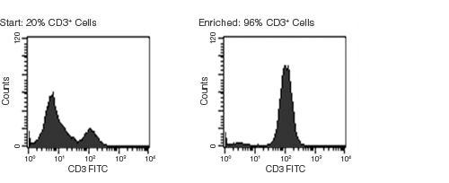
Typical RosetteSep™ HLA T Cell Enrichment Profile

Figure 1. Typical RosetteSep™ T Cell Enrichment Profile

Starting with fresh whole blood the CD3+ cell content of the enriched fraction typically ranges from 90% - 97%. Red blood cells were removed by lysis prior to flow cytometry.

Protocols and Documentation

Find supporting information and directions for use in the Product Information Sheet or explore additional protocols below.

Document Type
Product Name
Catalog #
Lot #
Language
Catalog #
15021, 15061
Lot #
All
Language
English
Document Type
Safety Data Sheet
Catalog #
15021, 15061
Lot #
All
Language
English

Applications

This product is designed for use in the following research area(s) as part of the highlighted workflow stage(s). Explore these workflows to learn more about the other products we offer to support each research area.

Resources and Publications

Frequently Asked Questions

What is RosetteSep™?

RosetteSep™ is a rapid cell separation procedure for the isolation of purified cells directly from whole blood, without columns or magnets.

How does RosetteSep™ work?

The antibody cocktail crosslinks unwanted cells to red blood cells (RBCs), forming rosettes. The unwanted cells then pellet with the free RBCs when centrifuged over a density centrifugation medium (e.g. Ficoll-Paque™ PLUS, Lymphoprep™).

What factors affect cell recovery?

The temperature of the reagents can affect cell recovery. All reagents should be at room temperature (sample, density centrifugation medium, PBS, centrifuge) before performing the isolations. Layering can also affect recovery so be sure to carefully layer the sample to avoid mixing with the density centrifugation medium as much as possible. Be sure to collect the entire enriched culture without disturbing the RBC pellet. A small amount of density centrifugation medium can be collected without worry.

Which cell samples can RosetteSep™ be used with?

RosetteSep™ can be used with leukapheresis samples, bone marrow or buffy coat, as long as: the concentration of cells does not exceed 5 x 10⁷ per mL (can dilute if necessary); and there are at least 100 RBCs for every nucleated cell (RBCs can be added if necessary).

Can RosetteSep™ be used with previously frozen or cultured cells?

Yes. Cells should be re-suspended at 2 - 5 x 10⁷ cells / mL in PBS + 2% FBS. Fresh whole blood should be added at 250 µL per mL of sample, as a source of red cells.

Can RosetteSep™ be used to enrich progenitors from cord blood?

Yes. Sometimes cord blood contains immature nucleated red cells that have a lower density than mature RBCs. These immature red cells do not pellet over Ficoll™, which can lead to a higher RBC contamination than peripheral blood separations.

Does RosetteSep™ work with mouse cells?

No, but we have developed EasySep™, a magnetic-based cell isolation system which works with mouse and other non-human species.

Which anticoagulant should be used with RosetteSep™?

Peripheral blood should be collected in heparinized Vacutainers. Cord blood should be collected in ACD.

Should the anticoagulant be washed off before using RosetteSep™?

No, the antibody cocktail can be added directly to the sample.

Publications (49)

PET Reporter Gene Imaging and Ganciclovir-Mediated Ablation of Chimeric Antigen Receptor T Cells in Solid Tumors. S. Murty et al. Cancer research 2020 nov

Abstract

Imaging strategies to monitor chimeric antigen receptor (CAR) T-cell biodistribution and proliferation harbor the potential to facilitate clinical translation for the treatment of both liquid and solid tumors. In addition, the potential adverse effects of CAR T cells highlight the need for mechanisms to modulate CAR T-cell activity. The herpes simplex virus type 1 thymidine kinase (HSV1-tk) gene has previously been translated as a PET reporter gene for imaging of T-cell trafficking in patients with brain tumor. The HSV1-TK enzyme can act as a suicide gene of transduced cells through treatment with the prodrug ganciclovir. Here we report the molecular engineering, imaging, and ganciclovir-mediated destruction of B7H3 CAR T cells incorporating a mutated version of the HSV1-tk gene (sr39tk) with improved enzymatic activity for ganciclovir. The sr39tk gene did not affect B7H3 CAR T-cell functionality and in vitro and in vivo studies in osteosarcoma models showed no significant effect on B7H3 CAR T-cell antitumor activity. PET/CT imaging with 9-(4-[18F]-fluoro-3-[hydroxymethyl]butyl)guanine ([18F]FHBG) of B7H3-sr39tk CAR T cells in an orthotopic model of osteosarcoma revealed tumor homing and systemic immune expansion. Bioluminescence and PET imaging of B7H3-sr39tk CAR T cells confirmed complete tumor ablation with intraperitoneal ganciclovir administration. This imaging and suicide ablation system can provide insight into CAR T-cell migration and proliferation during clinical trials while serving as a suicide switch to limit potential toxicities. SIGNIFICANCE: This study showcases the only genetically engineered system capable of serving the dual role both as an effective PET imaging reporter and as a suicide switch for CAR T cells.
A Simple and Scalable Strategy for Analysis of Endogenous Protein Dynamics. M. K. Schwinn et al. Scientific reports 2020 jun

Abstract

The ability to analyze protein function in a native context is central to understanding cellular physiology. This study explores whether tagging endogenous proteins with a reporter is a scalable strategy for generating cell models that accurately quantitate protein dynamics. Specifically, it investigates whether CRISPR-mediated integration of the HiBiT luminescent peptide tag can easily be accomplished on a large-scale and whether integrated reporter faithfully represents target biology. For this purpose, a large set of proteins representing diverse structures and functions, some of which are known or potential drug targets, were targeted for tagging with HiBiT in multiple cell lines. Successful insertion was detected for 86{\%} of the targets, as determined by luminescence-based plate assays, blotting, and imaging. In order to determine whether endogenously tagged proteins yield more representative models, cells expressing HiBiT protein fusions either from endogenous loci or plasmids were directly compared in functional assays. In the tested cases, only the edited lines were capable of accurately reproducing the anticipated biology. This study provides evidence that cell lines expressing HiBiT fusions from endogenous loci can be rapidly generated for many different proteins and that these cellular models provide insight into protein function that may be unobtainable using overexpression-based approaches.
PD-1+ melanocortin receptor dependent-Treg cells prevent autoimmune disease. F. Muhammad et al. Scientific reports 2019 nov

Abstract

Experimental autoimmune uveoretinitis (EAU) is a mouse model of human autoimmune uveitis marked by ocular autoantigen-specific regulatory immunity in the spleen. The melanocortin 5 receptor (MC5r) and adenosine 2 A receptor (A2Ar) are required for induction of post-EAU regulatory T cells (Tregs) which provide resistance to EAU. We show that blocking the PD-1/PD-L1 pathway prevented suppression of EAU by post-EAU Tregs. A2Ar induction of PD-1+FoxP3+ Tregs in uveitis patients was similar compared to healthy controls, but was significantly reduced with melanocortin stimulation. Further, lower body mass index correlated with responsiveness to stimulation of this pathway. These observations indicate an importance of the PD-1/PD-L1 pathway to provide resistance to relapsing uveitis and shows a reduced capacity of uveitis patients to induce Tregs when stimulated through melanocortin receptors, but that it is possible to bypass this part of the pathway through direct stimulation of A2Ar.
New look, same high quality and support! You may notice that your instrument or reagent packaging looks slightly different from images displayed on the website, or from previous orders. We are updating our look but rest assured, the products themselves and how you should use them have not changed. Learn more