RosetteSep™ Human NK Cell Enrichment Cocktail

Immunodensity negative selection cocktail

RosetteSep™ Human NK Cell Enrichment Cocktail

Immunodensity negative selection cocktail
From: 263 USD
Catalog #
(Select a product)
Immunodensity negative selection cocktail
Add to Wish List

Product Advantages


  • Fast and easy-to-use

  • Requires no special equipment or training

  • Isolated cells are untouched

  • Can be combined with SepMate™ for consistent, high-throughput sample processing

What's Included

  • RosetteSep™ Human NK Cell Enrichment Cocktail (Catalog #15025)
    • RosetteSep™ Human NK Cell Enrichment Cocktail, 2 mL
  • RosetteSep™ Human NK Cell Enrichment Cocktail (Catalog #15065)
    • RosetteSep™ Human NK Cell Enrichment Cocktail, 5 x 2 mL
Products for Your Protocol

Overview

The RosetteSep™ Human NK Cell Enrichment Cocktail is designed to isolate NK cells from whole blood by negative selection. Unwanted cells are targeted for removal with Tetrameric Antibody Complexes recognizing non-NK cells and red blood cells (RBCs). When centrifuged over a buoyant density medium such as RosetteSep™ DM-L (Catalog #15705) or Lymphoprep™ (Catalog #18060), the unwanted cells pellet along with the RBCs. The purified NK cells are present as a highly enriched population at the interface between the plasma and the buoyant density medium.
Subtype
Cell Isolation Kits
Cell Type
NK Cells
Species
Human
Sample Source
Buffy Coat, Whole Blood
Selection Method
Negative
Application
Cell Isolation
Brand
RosetteSep
Area of Interest
Immunology

Data Figures

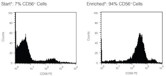
FACS Histogram Results Using RosetteSep™ Human NK Cell Enrichment Cocktail

Figure 1. FACS Histogram Results Using RosetteSep™ Human NK Cell Enrichment Cocktail

Starting with fresh peripheral blood the CD56+ cell content of the enriched fraction typically ranges from 80 - 98%. *Note: Red blood cells were removed by lysis prior to flow cytometry.

Protocols and Documentation

Find supporting information and directions for use in the Product Information Sheet or explore additional protocols below.

Document Type
Product Name
Catalog #
Lot #
Language
Catalog #
15065, 15025
Lot #
All
Language
English
Document Type
Safety Data Sheet
Catalog #
15065, 15025
Lot #
All
Language
English

Applications

This product is designed for use in the following research area(s) as part of the highlighted workflow stage(s). Explore these workflows to learn more about the other products we offer to support each research area.

Resources and Publications

Frequently Asked Questions

What is RosetteSep™?

RosetteSep™ is a rapid cell separation procedure for the isolation of purified cells directly from whole blood, without columns or magnets.

How does RosetteSep™ work?

The antibody cocktail crosslinks unwanted cells to red blood cells (RBCs), forming rosettes. The unwanted cells then pellet with the free RBCs when centrifuged over a density centrifugation medium (e.g. Ficoll-Paque™ PLUS, Lymphoprep™).

What factors affect cell recovery?

The temperature of the reagents can affect cell recovery. All reagents should be at room temperature (sample, density centrifugation medium, PBS, centrifuge) before performing the isolations. Layering can also affect recovery so be sure to carefully layer the sample to avoid mixing with the density centrifugation medium as much as possible. Be sure to collect the entire enriched culture without disturbing the RBC pellet. A small amount of density centrifugation medium can be collected without worry.

Which cell samples can RosetteSep™ be used with?

RosetteSep™ can be used with leukapheresis samples, bone marrow or buffy coat, as long as: the concentration of cells does not exceed 5 x 10⁷ per mL (can dilute if necessary); and there are at least 100 RBCs for every nucleated cell (RBCs can be added if necessary).

Can RosetteSep™ be used with previously frozen or cultured cells?

Yes. Cells should be re-suspended at 2 - 5 x 10⁷ cells / mL in PBS + 2% FBS. Fresh whole blood should be added at 250 µL per mL of sample, as a source of red cells.

Can RosetteSep™ be used to enrich progenitors from cord blood?

Yes. Sometimes cord blood contains immature nucleated red cells that have a lower density than mature RBCs. These immature red cells do not pellet over Ficoll™, which can lead to a higher RBC contamination than peripheral blood separations.

Does RosetteSep™ work with mouse cells?

No, but we have developed EasySep™, a magnetic-based cell isolation system which works with mouse and other non-human species.

Which anticoagulant should be used with RosetteSep™?

Peripheral blood should be collected in heparinized Vacutainers. Cord blood should be collected in ACD.

Should the anticoagulant be washed off before using RosetteSep™?

No, the antibody cocktail can be added directly to the sample.

Publications (71)

Identification of elements determining KIR gene demethylation at the CD56-bright stage of NK cell development. H. Li et al. iScience 2026 Sep

Abstract

The variegated expression of the KIR family of class I MHC receptors generates specialized natural killer (NK) cells capable of allele-specific HLA recognition. Understanding the mechanism of KIR gene activation will lead to improved methods for the generation of fully functional NK cells. A central RUNX-binding site in the KIR proximal promoter is required for gene activation. RUNX proteins recruit ten-eleven translocation (TET) proteins that generate 5-hydroxymethylcytosine (5hmC) and drive DNA demethylation. Assessment of 5-methylcytosine (5mC) and 5hmC residues at four stages of NK cell development reveals deposition of 5hmC primarily in a CREB site next to the RUNX site at the CD56Bright stage but not the subsequent CD56Dim stage representing fully mature NK cells. KIR promoter demethylation is delayed relative to other lineage-associated genes, indicating a high threshold for KIR gene demethylation in developing NK cells, and a window of opportunity for RUNX/TET-dependent KIR gene activation in CD56Bright NK cells.
Longitudinal assessment of functional antibodies to a novel influenza virus strain across age groups. A. Huckriede et al. PLoS pathogens 2026 Sep

Abstract

Newly emerging influenza viruses pose a constant threat as they encounter a population lacking neutralizing antibodies against the new strain. However, cross-reactive non-neutralizing antibodies (nnABs) may be present and help mitigate disease symptoms through various effector mechanisms, including antibody-dependent cellular cytotoxicity (ADCC). Although nnABs to influenza virus have received more attention lately, little information is available on their age-related prevalence, steady-state levels, functional properties, and changes in these parameters over time. Using longitudinal samples from adolescents, adults, and older adults, collected before and after the 2009 swine flu pandemic, we comprehensively characterized the specificity and functionality of nnAB responses against H1N1 pandemic 2009 (H1N1pdm09) virus. Remarkably, all participants exhibited cross-reactive antibodies to this virus before having encountered it through infection or vaccination, with the highest baseline levels observed in older adults. The levels of these IgG antibodies showed a strong correlation with engagement of fragment crystallizable γ receptor IIIa (FcγRIIIa) and NK cell activation, both of which were notably lower in adolescents than in adults and older adults. Without infection or vaccination, average amounts of H1N1pdm09-reactive antibodies remained relatively stable on the population level over the 5-year study period. However, on an individual level, substantial increases and decreases occurred. H1N1pdm09 infection or vaccination significantly enhanced specific antibody levels and the FcγRIIIa-engaging capacity of these antibodies in all age groups. ADCC-mediating antibodies increased, however, only in adolescents, reaching the same level as observed in the adult groups. Taken together, our results demonstrate the presence of cross-reactive, non-neutralizing, functional, and boostable antibodies against a never-encountered influenza virus strain across all age groups. These antibodies might contribute to protection from severe disease. Accordingly, in case of a newly emerging influenza virus, their further enhancement by vaccination could be beneficial as an immediate protective measure before a strain-specific vaccine becomes available.
Rituximab Binding Endows CD20+ Extracellular Vesicles With NK Cell-Activating Properties in B Cell Lymphoma. A. Otero et al. Journal of extracellular vesicles 2026 Aug

Abstract

Non-Hodgkin lymphoma (NHL), predominantly B cell lymphomas (B-NHL), is currently treated with chemotherapy combined with rituximab (RTX), an anti-CD20 monoclonal antibody. Despite substantial therapeutic advances, treatment resistance and disease relapse continue to affect a significant fraction of patients. The mechanisms by which the tumour microenvironment and other factors influence RTX efficacy are not fully elucidated. Herein, we hypothesized that CD20+ extracellular vesicles (EVs) shed by B cell lymphomas are recognized by RTX forming immune complexes that modulate natural killer (NK) cell activity via Fcγ receptor (FcγR) interactions. EVs isolated from lymph node explants and plasma samples of B-NHL patients contained abundant CD20+ vesicles, which were particularly enriched in advanced disease. RTX specifically bound CD20 on these EVs, generating EV-RTX immune complexes. Functional studies employing EVs from a B-NHL cell line and from patient-derived samples demonstrated that, whereas EVs suppressed NK cell activation and cytotoxicity, EV-RTX immune complexes reversed this inhibitory effect and enhanced NK cell effector functions. Indeed, EV-RTX immune complexes specifically triggered FcγRIIIa-dependent NK cell activation, evidenced by increased Syk phosphorylation, CD69 expression, and enhanced lytic activity against target cells. Our findings uncover a previously unrecognized mechanism by which RTX, through the formation of immune complexes with CD20+ EVs, promotes NK cell activation and may enhance therapeutic efficacy. More broadly, these results demonstrate that antibody binding can endow EVs with novel immunomodulatory properties, revealing a potential mechanism by which therapeutic antibodies reshape EV function and influence anti-tumour immunity.