MesenCult™ Adipogenic Differentiation Kit (Mouse)

For the in vitro differentiation of mouse MSCs, ADSCs, and MEFs into adipocytes

MesenCult™ Adipogenic Differentiation Kit (Mouse)

For the in vitro differentiation of mouse MSCs, ADSCs, and MEFs into adipocytes

From: 316 USD
Catalog #
(Select a product)
For the in vitro differentiation of mouse MSCs, ADSCs, and MEFs into adipocytes
Add to Wish List

Product Advantages


  • Compatible with mouse MSCs previously culture-expanded using the MesenCult™ Expansion Kit (Mouse).

  • Available in an easy-to-use two-component format.

  • Rigorous raw material screening and quality control minimize lot-to-lot variablity.

What's Included

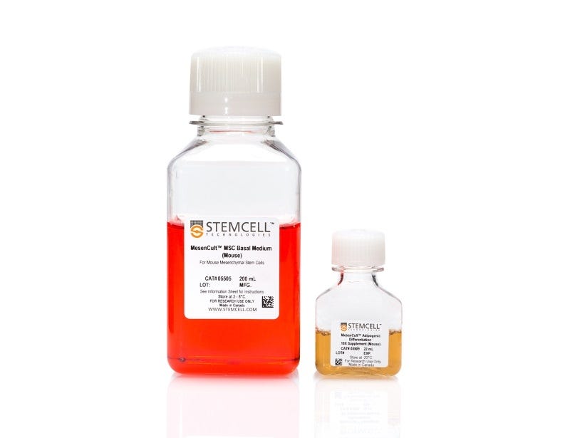
  • MesenCult™ MSC Basal Medium (Mouse), 200 mL
  • MesenCult™ Adipogenic Differentiation 10X Supplement (Mouse), 22 mL

Overview

MesenCult™ Adipogenic Differentiation Kit (Mouse) is specifically formulated for the in vitro differentiation of mouse mesenchymal stem and progenitor cells (MSCs), adipose tissue-derived MSCs (ADSCs), and mouse embryonic fibroblasts (MEFs) into cells of the adipogenic lineage.
NOTE: MesenCult™ Adipogenic Differentiation Medium must be supplemented with L-Glutamine (Catalog #07100).
Subtype
Specialized Media
Cell Type
Mesenchymal Stem and Progenitor Cells
Species
Mouse
Application
Differentiation
Brand
MesenCult
Area of Interest
Stem Cell Biology

Protocols and Documentation

Find supporting information and directions for use in the Product Information Sheet or explore additional protocols below.

Document Type
Product Name
Catalog #
Lot #
Language
Catalog #
05507
Lot #
All
Language
English
Document Type
Safety Data Sheet 1
Catalog #
05507
Lot #
All
Language
English
Document Type
Safety Data Sheet 2
Catalog #
05507
Lot #
All
Language
English

Applications

This product is designed for use in the following research area(s) as part of the highlighted workflow stage(s). Explore these workflows to learn more about the other products we offer to support each research area.

Resources and Publications

Publications (5)

Site-1 protease ablation in the osterix-lineage in mice results in bone marrow neutrophilia and hematopoietic stem cell alterations. D. Patra et al. Biology open 2020 jun

Abstract

Site-1 protease (S1P) ablation in the osterix-lineage in mice drastically reduces bone development and downregulates bone marrow-derived skeletal stem cells. Here we show that these mice also suffer from spina bifida occulta with a characteristic lack of bone fusion in the posterior neural arches. Molecular analysis of bone marrow-derived non-red blood cell cells, via single-cell RNA-Seq and protein mass spectrometry, demonstrate that these mice have a much-altered bone marrow with a significant increase in neutrophils and Ly6C-expressing leukocytes. The molecular composition of bone marrow neutrophils is also different as they express more and additional members of the stefin A (Stfa) family of proteins. In vitro, recombinant Stfa1 and Stfa2 proteins have the ability to drastically inhibit osteogenic differentiation of bone marrow stromal cells, with no effect on adipogenic differentiation. FACS analysis of hematopoietic stem cells show that despite a decrease in hematopoietic stem cells, S1P ablation results in an increased production of granulocyte-macrophage progenitors, the precursors to neutrophils. These observations indicate that S1P has a role in the lineage specification of hematopoietic stem cells and/or their progenitors for development of a normal hematopoietic niche. Our study designates a fundamental requirement of S1P for maintaining a balanced regenerative capacity of the bone marrow niche.
3D-printable supramolecular hydrogels with shear-thinning property: fabricating strength tunable bioink via dual crosslinking. T. Hu et al. Bioactive materials 2020 dec

Abstract

3-dimensional (3D) bioprinting technology provides promising strategy in the fabrication of artificial tissues and organs. As the fundamental element in bioprinting process, preparation of bioink with ideal mechanical properties without sacrifice of biocompatibility is a great challenge. In this study, a supramolecular hydrogel-based bioink is prepared by polyethylene glycol (PEG) grafted chitosan, $\alpha$-cyclodextrin ($\alpha$-CD) and gelatin. It has a primary crosslinking structure through the aggregation of the pseudo-polyrotaxane-like side chains, which are formed from the host-guest interactions between $\alpha$-CD and PEG side chain. Apparent viscosity measurement shows the shear-shinning property of this bioink, which might be due to the reversibility of the physical crosslinking. Moreover, with $\beta$-glycerophosphate at different concentrations as the secondary crosslinking agent, the printed constructs demonstrate different Young's modulus (p {\textless} 0.001). They could also maintain the Young's modulus in cell culture condition for at least 21 days (p {\textless} 0.05). By co-culturing each component with fibroblasts, CCK-8 assay demonstrate cellular viability is higher than 80{\%}. After bioprinting and culturing, immunofluorescence staining with quantification indicate the expression of Ki-67, Paxillin, and N-cadherin is higher in day 14 than those in day 3 (p {\textless} 0.05). Oil red O and Nissl body specific staining reflect strength tunable bioink may have impact on the cell fate of mesenchymal stem cells (p {\textless} 0.05). This work might provide new idea for advanced bioink in the application of re-establishing complicated tissues and organs.
Machine Learning to Quantitate Neutrophil NETosis. L. Elsherif et al. Scientific reports 2019 nov

Abstract

We introduce machine learning (ML) to perform classification and quantitation of images of nuclei from human blood neutrophils. Here we assessed the use of convolutional neural networks (CNNs) using free, open source software to accurately quantitate neutrophil NETosis, a recently discovered process involved in multiple human diseases. CNNs achieved {\textgreater}94{\%} in performance accuracy in differentiating NETotic from non-NETotic cells and vastly facilitated dose-response analysis and screening of the NETotic response in neutrophils from patients. Using only features learned from nuclear morphology, CNNs can distinguish between NETosis and necrosis and between distinct NETosis signaling pathways, making them a precise tool for NETosis detection. Furthermore, by using CNNs and tools to determine object dispersion, we uncovered differences in NETotic nuclei clustering between major NETosis pathways that is useful in understanding NETosis signaling events. Our study also shows that neutrophils from patients with sickle cell disease were unresponsive to one of two major NETosis pathways. Thus, we demonstrate the design, performance, and implementation of ML tools for rapid quantitative and qualitative cell analysis in basic science.