EpiCult™-B Mouse Medium Kit

For culture and evaluation of mouse mammary epithelial cells

EpiCult™-B Mouse Medium Kit

For culture and evaluation of mouse mammary epithelial cells

From: 353 USD
Catalog #
(Select a product)
For culture and evaluation of mouse mammary epithelial cells
Add to Wish List

What's Included

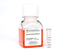
  • EpiCult™-B Basal Medium (Mouse), 450 mL
  • EpiCult™-B Proliferation Supplement (Mouse), 50 mL
Products for Your Protocol
To see all required products for your protocol, please consult the Protocols and Documentation.

Overview

EpiCult™-B (mouse) is a serum-free liquid culture medium optimized for the culture of mouse mammary luminal and myoepithelial cells. It is ideal for the culture and evaluation of mouse mammary epithelial progenitors in the mammary colony-forming unit assay when used in conjunction with an irradiated feeder layer such as NIH 3T3 cells. This medium is also used for enzymatic dissociation of mouse mammary tissue when supplemented with collagenase and hyaluronidase. Addition of Human Recombinant EGF (Catalog #78006), Human Recombinant bFGF (Catalog #78003), and Heparin Solution (Catalog #07980) is required for culturing cells.
Subtype
Specialized Media
Cell Type
Mammary Cells
Species
Mouse
Application
Cell Culture, Colony Assay
Brand
EpiCult
Area of Interest
Epithelial Cell Biology
Formulation Category
Serum-Free

Data Figures

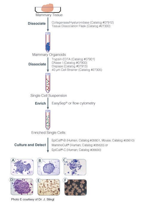
Protocol for isolation and identification of human and mouse mammary epithelial progenitor cells

Figure 1. Protocol for Isolation and Identification of Human and Mouse Mammary Epithelial Progenitor Cells

Phase contrast photographs of (A) a pure human myoepithelial cell colony, (B) a pure human luminal cell colony, and (C) a mixed human colony. (D) is a mouse colony. Unlike human mammary CFC colonies, subtypes of mouse mammary epithelial cell colonies are not easily identifiable. All colonies were cultured in either EpiCult®-B (Human: Catalog #05601) or EpiCult®-B (Mouse:Catalog #5610) in the presence of an irradiated NIH 3T3 feeder layer. Colonies were visualized by staining with Wright"s Giemsa. (E) is a picture of mammospheres obtained from primary human mammary epithelial cells and (F) is an image of tumorspheres obtained from MCF7 human breast cancer cell line.

Protocols and Documentation

Find supporting information and directions for use in the Product Information Sheet or explore additional protocols below.

Document Type
Product Name
Catalog #
Lot #
Language
Catalog #
05610
Lot #
All
Language
English
Document Type
Safety Data Sheet 1
Catalog #
05610
Lot #
All
Language
English
Document Type
Safety Data Sheet 2
Catalog #
05610
Lot #
All
Language
English

Applications

This product is designed for use in the following research area(s) as part of the highlighted workflow stage(s). Explore these workflows to learn more about the other products we offer to support each research area.

Resources and Publications

Publications (9)

Sterol regulatory element binding protein 1 couples mechanical cues and lipid metabolism. R. Bertolio et al. Nature communications 2019

Abstract

Sterol regulatory element binding proteins (SREBPs) are a family of transcription factors that regulate lipid biosynthesis and adipogenesis by controlling the expression of several enzymes required for cholesterol, fatty acid, triacylglycerol and phospholipid synthesis. In vertebrates, SREBP activation is mainly controlled by a complex and well-characterized feedback mechanism mediated by cholesterol, a crucial bio-product of the SREBP-activated mevalonate pathway. In this work, we identified acto-myosin contractility and mechanical forces imposed by the extracellular matrix (ECM) as SREBP1 regulators. SREBP1 control by mechanical cues depends on geranylgeranyl pyrophosphate, another key bio-product of the mevalonate pathway, and impacts on stem cell fate in mouse and on fat storage in Drosophila. Mechanistically, we show that activation of AMP-activated protein kinase (AMPK) by ECM stiffening and geranylgeranylated RhoA-dependent acto-myosin contraction inhibits SREBP1 activation. Our results unveil an unpredicted and evolutionary conserved role of SREBP1 in rewiring cell metabolism in response to mechanical cues.
Development of mammary luminal progenitor cells is controlled by the transcription factor STAT5A. Yamaji D et al. Genes & development 2009 OCT

Abstract

Mammary alveologenesis is abrogated in the absence of the transcription factors STAT5A/5B, which mediate cytokine signaling. To reveal the underlying causes for this developmental block, we studied mammary stem and progenitor cells. While loss of STAT5A/5B did not affect the stem cell population and its ability to form mammary ducts, luminal progenitors were greatly reduced and unable to form alveoli during pregnancy. Temporally controlled expression of transgenic STAT5A in mammary epithelium lacking STAT5A/5B restored the luminal progenitor population and rescued alveologenesis in a reversible fashion in vivo. Thus, STAT5A is necessary and sufficient for the establishment of luminal progenitor cells.
MMTV-Wnt1 and -DeltaN89beta-catenin induce canonical signaling in distinct progenitors and differentially activate Hedgehog signaling within mammary tumors. Teissedre B et al. PloS one 2009 JAN

Abstract

Canonical Wnt/beta-catenin signaling regulates stem/progenitor cells and, when perturbed, induces many human cancers. A significant proportion of human breast cancer is associated with loss of secreted Wnt antagonists and mice expressing MMTV-Wnt1 and MMTV-DeltaN89beta-catenin develop mammary adenocarcinomas. Many studies have assumed these mouse models of breast cancer to be equivalent. Here we show that MMTV-Wnt1 and MMTV-DeltaN89beta-catenin transgenes induce tumors with different phenotypes. Using axin2/conductin reporter genes we show that MMTV-Wnt1 and MMTV-DeltaN89beta-catenin activate canonical Wnt signaling within distinct cell-types. DeltaN89beta-catenin activated signaling within a luminal subpopulation scattered along ducts that exhibited a K18(+)ER(-)PR(-)CD24(high)CD49f(low) profile and progenitor properties. In contrast, MMTV-Wnt1 induced canonical signaling in K14(+) basal cells with CD24/CD49f profiles characteristic of two distinct stem/progenitor cell-types. MMTV-Wnt1 produced additional profound effects on multiple cell-types that correlated with focal activation of the Hedgehog pathway. We document that large melanocytic nevi are a hitherto unreported hallmark of early hyperplastic Wnt1 glands. These nevi formed along the primary mammary ducts and were associated with Hedgehog pathway activity within a subset of melanocytes and surrounding stroma. Hh pathway activity also occurred within tumor-associated stromal and K14(+)/p63(+) subpopulations in a manner correlated with Wnt1 tumor onset. These data show MMTV-Wnt1 and MMTV-DeltaN89beta-catenin induce canonical signaling in distinct progenitors and that Hedgehog pathway activation is linked to melanocytic nevi and mammary tumor onset arising from excess Wnt1 ligand. They further suggest that Hedgehog pathway activation maybe a critical component and useful indicator of breast tumors arising from unopposed Wnt1 ligand.