MethoCult™ M3234

Methylcellulose-based medium without cytokines (including EPO) for mouse cells

MethoCult™ M3234

Methylcellulose-based medium without cytokines (including EPO) for mouse cells

From: 414 USD
Catalog #
(Select a product)
Methylcellulose-based medium without cytokines (including EPO) for mouse cells
Add to Wish List

Overview

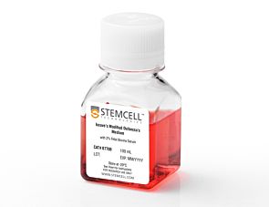
MethoCult™ M3234 is an incomplete medium formulation that contains methylcellulose in Iscove's MDM. MethoCult™ M3234 is suitable for the growth and enumeration of hematopoietic progenitor cells in colony-forming unit (CFU) assays of mouse bone marrow, spleen, peripheral blood, and fetal liver, when the appropriate growth factors and supplements are added. This formulation does not contain erythropoietin or other cytokines.

Browse our Frequently Asked Questions (FAQs) on performing the CFU assay.
Contains
• Methylcellulose in Iscove's MDM
• Fetal bovine serum
• Bovine serum albumin
• Recombinant human insulin
• Human transferrin (iron-saturated)
• 2-Mercaptoethanol
• Supplements
Subtype
Semi-Solid Media, Specialized Media
Cell Type
Hematopoietic Stem and Progenitor Cells
Species
Mouse
Application
Cell Culture, Colony Assay, Functional Assay
Brand
MethoCult
Area of Interest
Stem Cell Biology
Formulation Category
Methylcellulose-Based

Protocols and Documentation

Find supporting information and directions for use in the Product Information Sheet or explore additional protocols below.

Document Type
Product Name
Catalog #
Lot #
Language
Catalog #
03234
Lot #
All
Language
English
Document Type
Technical Manual
Catalog #
03234
Lot #
All
Language
English
Document Type
Safety Data Sheet
Catalog #
03234
Lot #
All
Language
English

Applications

This product is designed for use in the following research area(s) as part of the highlighted workflow stage(s). Explore these workflows to learn more about the other products we offer to support each research area.

Resources and Publications

Frequently Asked Questions

Why use semi-solid media?

Semi-solid media (methylcellulose-based MethoCult™ and collagen-based MegaCult™-C) allow the clonal progeny of a single progenitor cell to remain spatially isolated from other colonies within a culture, so they may be separately identified and counted.

Why use methylcellulose-based media?

Methylcellulose permits better growth of erythroid colonies than other types of semi-solid support systems (eg. agar) while allowing optimal myeloid colony formation. When appropriate cytokines are present, committed progenitor cells of both erythroid and granulocyte/macrophage lineages (CFU-GM, CFU-G, CFU-M) as well as multi-potential progenitor cells (CFU-GEMM), can be assayed simultaneously in the same culture dish.

Is it necessary to add antibiotics to the media?

No, aseptic technique should be sufficient to maintain sterile cultures. However, antibiotics (eg. Penicillin/Streptomycin) or anti-fungals (eg. Amphotericin B) may be added to the methylcellulose medium if desired.

Is there anything I can do if my cultures appear contaminated?

No, once contamination is visible, it is not possible to rescue the cultures by the addition of antibiotics. Bacteria and yeast inhibit colony formation by depleting nutrients or by releasing toxic substances.

Why can't I use a pipette to dispense methylcellulose-based media?

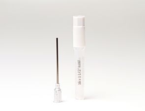
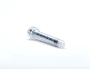
Methylcellulose is a viscous solution that cannot be accurately dispensed using a pipette due to adherence of the medium to the walls of the pipette tip. Blunt-End, 16 Gauge needles (Catalog #28110), in combination with 3 cc Syringes (Catalog #28230) are recommended for accurate dispensing of MethoCult™.

Can I 'pluck' the colonies for individual analysis?

Yes, colonies can be 'plucked' using a pipette with 200 µL sterile pipette tips or using a glass Pasteur pipette with an elongated tip. Individual colonies should be placed in a volume of 25 - 50 µL of medium, and diluted into suitable culture medium for further culture or analysis.

Why are low adherence dishes so important?

Adherent cells such as fibroblasts can cause inhibition of colony growth and obscure visualization of colonies.

Can MethoCult™ products be used for lymphoid progenitor CFU assays?

Human lymphoid progenitors (B, NK and T) seem to require stromal support for growth therefore cannot be grown in MethoCult™. Mouse pre-B clonogenic progenitors can be grown in MethoCult™ M3630 (Catalog #03630).

Is it possible to set up CFU assays in a 24-well plate?

Yes, as long as a plating concentration optimized for the smaller surface area of a well in a 24-well plate (1.9 cm2 as compared to ~9.5 cm2 for a 35 mm dish) is used for these assays. The number of replicate wells required to get an accurate estimation of CFU numbers may also need to be increased.

Can I stain colonies in MethoCult™ medium?

The cells in individual colonies in MethoCult™ can be stained, eg., for analysis of morphology or phenotype, after they are plucked from the dish and washed free of methylcellulose. Colonies grown in collagen-based MegaCult™-C medium can be used for immunohistochemical or enzymatic staining in situ after dehydration and fixation onto glass slides.

Are there differences in colony morphology with serum-free media?

Serum-containing media generally give better overall growth (colonies may appear larger) but there are no large differences in total colony numbers when CFU assays using serum-free media and serum-containing media are compared, provided that identical cytokines are present.

Can MethoCult™ be made with alternate base media?

Yes, this can be done as a 'custom' media order. Please contact techsupport@stemcell.com for more information.

Is there a MethoCult™ formulation suitable for HPP-CFC (high proliferative potential colony forming cell)?

Yes, MethoCult™ H4535 (Catalog #04535) can be used for the HPP-CFC assay as it does not contain EPO. The culture period is usually 28 days. It is not necessary to feed these cultures as growth factors in the medium are present in excess. As HPP-CFCs can be quite large, overplating can be a problem. It is recommended to plate cells at two or more different concentrations.

Publications (37)

Population snapshots predict early haematopoietic and erythroid hierarchies. B. K. Tusi et al. Nature 2018 FEB

Abstract

The formation of red blood cells begins with the differentiation of multipotent haematopoietic progenitors. Reconstructing the steps of this differentiation represents a general challenge in stem-cell biology. Here we used single-cell transcriptomics, fate assays and a theory that allows the prediction of cell fates from population snapshots to demonstrate that mouse haematopoietic progenitors differentiate through a continuous, hierarchical structure into seven blood lineages. We uncovered coupling between the erythroid and the basophil or mast cell fates, a global haematopoietic response to erythroid stress and novel growth factor receptors that regulate erythropoiesis. We defined a flow cytometry sorting strategy to purify early stages of erythroid differentiation, completely isolating classically defined burst-forming and colony-forming progenitors. We also found that the cell cycle is progressively remodelled during erythroid development and during a sharp transcriptional switch that ends the colony-forming progenitor stage and activates terminal differentiation. Our work showcases the utility of linking transcriptomic data to predictive fate models, and provides insights into lineage development in vivo.
Requirement for Dot1l in murine postnatal hematopoiesis and leukemogenesis by MLL translocation. Jo SY et al. Blood 2011 MAY

Abstract

Disruptor of telomeric silencing 1-like (Dot1l) is a histone 3 lysine 79 methyltransferase. Studies of constitutive Dot1l knockout mice show that Dot1l is essential for embryonic development and prenatal hematopoiesis. DOT1L also interacts with translocation partners of Mixed Lineage Leukemia (MLL) gene, which is commonly translocated in human leukemia. However, the requirement of Dot1l in postnatal hematopoiesis and leukemogenesis of MLL translocation proteins has not been conclusively shown. With a conditional Dot1l knockout mouse model, we examined the consequences of Dot1l loss in postnatal hematopoiesis and MLL translocation leukemia. Deletion of Dot1l led to pancytopenia and failure of hematopoietic homeostasis, and Dot1l-deficient cells minimally reconstituted recipient bone marrow in competitive transplantation experiments. In addition, MLL-AF9 cells required Dot1l for oncogenic transformation, whereas cells with other leukemic oncogenes, such as Hoxa9/Meis1 and E2A-HLF, did not. These findings illustrate a crucial role of Dot1l in normal hematopoiesis and leukemogenesis of specific oncogenes.
HIF1alpha synergizes with glucocorticoids to promote BFU-E progenitor self-renewal. Flygare J et al. Blood 2011 MAR

Abstract

With the aim of finding small molecules that stimulate erythropoiesis earlier than erythropoietin and that enhance erythroid colony-forming unit (CFU-E) production, we studied the mechanism by which glucocorticoids increase CFU-E formation. Using erythroid burst-forming unit (BFU-E) and CFU-E progenitors purified by a new technique, we demonstrate that glucocorticoids stimulate the earliest (BFU-E) progenitors to undergo limited self-renewal, which increases formation of CFU-E cells textgreater 20-fold. Interestingly, glucocorticoids induce expression of genes in BFU-E cells that contain promoter regions highly enriched for hypoxia-induced factor 1α (HIF1α) binding sites. This suggests activation of HIF1α may enhance or replace the effect of glucocorticoids on BFU-E self-renewal. Indeed, HIF1α activation by a prolyl hydroxylase inhibitor (PHI) synergizes with glucocorticoids and enhances production of CFU-Es 170-fold. Because PHIs are able to increase erythroblast production at very low concentrations of glucocorticoids, PHI-induced stimulation of BFU-E progenitors thus represents a conceptually new therapeutic window for treating erythropoietin-resistant anemia.