Thank you for your interest in this product.
Please provide us with your contact information and your local representative
will contact you with a customized quote. Where appropriate, they can also assist you with a(n):
Estimated delivery time for your area
Product sample or exclusive offer
In-lab demonstration
By submitting this form, you are providing your consent to STEMCELL Technologies Canada Inc. and its subsidiaries and affiliates (“STEMCELL”) to collect and use your information, and send you newsletters and emails in accordance with our privacy policy. Please contact us with any questions that you may have. You can unsubscribe or change your email preferences at any time.
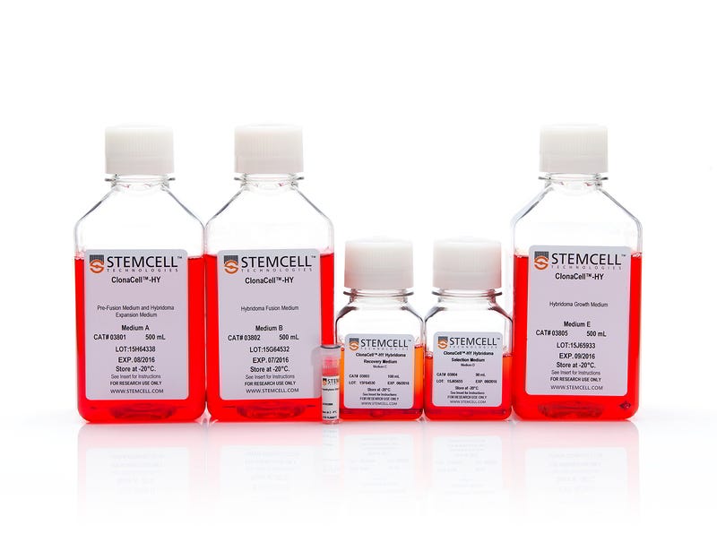
The ClonaCell™-HY method uses a methylcellulose-based semi-solid selective medium to combine hybridoma selection and cloning into one step. Individual parental hybridoma clones and their progeny remain localized together in the semi-solid medium as they grow to form distinct colonies. This prevents the loss of rare clones by overgrowth from faster-growing cells, as can occur during selection in a liquid medium. The hybridoma colonies can be easily picked from the semi-solid medium by manual or robotic methods and dispersed into a liquid growth medium for screening and expansion.
This kit has been verified for use in mouse and rat hydridoma development and monoclonal antibody production and reportedly is compatible for production, cloning, and expansion of hybridomas using lymphocytes from a variety of host animals including human, mouse, rat, and hamster. For your convenience, the kit components are also available for purchase individually.
Subtype
Semi-Solid Media, Specialized Media
Cell Type
Hybridomas
Species
Mouse, Other, Rat
Application
Cell Culture, Hybridoma Generation
Brand
ClonaCell
Area of Interest
Antibody Development, Cell Line Development, Drug Discovery and Toxicity Testing, Hybridoma Generation
This product is designed for use in the following research area(s) as part
of the highlighted workflow stage(s). Explore these workflows to learn more about the other products we
offer to support each research area.
Why is there HT (hypoxanthine, thymidine) in Medium E?
Hybridomas are selected using HAT (hypoxanthine, aminopterin, thymidine). Aminopterin blocks the de novo pathway for synthesizing nucleotide precursors for DNA synthesis. The inhibition of the de novo pathway can persist even after the cells are removed from selection. Hypoxanthine and thymidine (HT) provide the necessary nucleotide precursors for hybridoma cells to synthesize DNA using the salvage pathway. Once the cells are growing well in Medium E, they can be gradually switched to Medium A or another medium without HT.
Is the serum in ClonaCell™-HY media heat inactivated?
Yes, all serum used in ClonaCell™-HY media is heat inactivated.
Is there any IgG in clonacell™-HY media?
While we don't add IgG to the ClonaCell™-HY media, we do add serum, which contains an undefined amount of IgG. We selectively use serum lots with low IgG levels in the production of ClonaCell™-HY media, however, levels vary from lot to lot. IgG levels in a specific lot of ClonaCell™-HY medium are available in the lot-specific Certificate of Analysis.
Are there antibiotics in ClonaCell™-HY media?
These products contain gentamycin rather than penicillin/streptomycin/amphotericin B, because gentamycin is more stable and is a broad spectrum antibiotic that is non-toxic to most mammalian cells in culture.
What is the optimal number of colonies per plate?
We recommend 50-150 colonies per plate. An average fusion will result in approximately 1000 colonies per fusion (approx. 100 colonies per plate). Even if the average number of colonies per plate approaches 300, there should still be enough separation between colonies to pick easily.
Why do I have to put my fused cells into liquid medium overnight? Why can't I just plate directly into Medium D?
We recommend waiting up to 24 hours so that all of the fused cells can go through one cell cycle. This will ensure they have a chance to express HPRT (hypoxanthine guanine phosphoribosyltransferase), the enzyme necessary to survive in the presence of aminopterin (present in Medium D). Additionally, fused cells are very fragile immediately after fusion. Waiting a day before mixing the cells with the methylcellulose will improve their survival. Although it is not recommended, fused cells may be plated on the same day as fusion, but the cells should be allowed to recover for several hours in ClonaCell™-HY Medium C prior to plating.
What myeloma and mouse strains should I use?
Myeloma: There are at least two common myeloma cell lines used to generate hybridomas - SP2/0 and P3X63Ag8.683. Both are available from ATCC. Researchers should ensure that the myeloma line is from a reliable source and is negative for mycoplasma. Mycoplasma contamination of the myeloma line can result in decreased efficiency of hybridoma formation. Mouse: We suggest using BALB/c splenocytes and parental myeloma cells of BALB/c for the following reasons: they are highly immune reactive, well characterized and myeloma cells are available from the same genetic strain. Other mouse strains, however, are also compatible with cloning in ClonaCell™-HY media.
Can I grow human/rat/T cell hybridomas in ClonaCell™-HY?
Although we have not tried to generate human, rat or T cell hybridomas during in-house testing, these experiments are expected to be successful using ClonaCell™-HY. The researcher would need to ensure that the cell lines used in the fusion are sensitive to HAT selection and grow well in methylcellulose-based medium.
There are very few colonies growing in my Medium D. Why?
Low numbers of colonies is generally a result of low fusion efficiency, which can have many causes. The fusion efficiency can be affected by the presence of serum during fusion, the presence of mycoplasma, low viability of cells, overexposure to polyethylene glycol or slow-growing myeloma cells prior to fusion.
Why does the ClonaCell™-HY manual suggest two different methods for fusion (A or B)? Can one expect better results with one method over the other?
Which method chosen is a personal preference and there should not be significant differences in efficiency. Method B is faster and has less steps, but Method B requires you to remove all the PEG before the cells are diluted, so you will risk aspirating cells if not very careful. With Method A, you dilute the PEG with Medium B, so you have less opportunity to lose cells.
Why does the ClonaCell™-HY manual suggest two different methods for fusion (A or B)? Can one expect better results with one method over the other?
A: Which method chosen is a personal preference and there should not be significant differences in efficiency. Method B is faster and has less steps, but Method B requires you to remove all the PEG before the cells are diluted, so you will risk aspirating cells if not very careful. With Method A, you dilute the PEG with Medium B, so you have less opportunity to lose cells.
Once I pick the colonies and grow the cells in plates, will the residual methylcellulose interfere with characterization? For example, will I have problems doing an ELISA?
There will likely be some residual methylcellulose contamination when colonies are picked and transferred to the 96-well plate with the liquid growth medium. The concentration of methylcellulose, however, should be low enough that it should not interfere with most assays.
Structural basis for nonneutralizing antibody competition at antigenic site II of the respiratory syncytial virus fusion protein.
Mousa JJ et al.
Proceedings of the National Academy of Sciences of the United States of America 2016 OCT
Abstract
Palivizumab was the first antiviral monoclonal antibody (mAb) approved for therapeutic use in humans, and remains a prophylactic treatment for infants at risk for severe disease because of respiratory syncytial virus (RSV). Palivizumab is an engineered humanized version of a murine mAb targeting antigenic site II of the RSV fusion (F) protein, a key target in vaccine development. There are limited reported naturally occurring human mAbs to site II; therefore, the structural basis for human antibody recognition of this major antigenic site is poorly understood. Here, we describe a nonneutralizing class of site II-specific mAbs that competed for binding with palivizumab to postfusion RSV F protein. We also describe two classes of site II-specific neutralizing mAbs, one of which escaped competition with nonneutralizing mAbs. An X-ray crystal structure of the neutralizing mAb 14N4 in complex with F protein showed that the binding angle at which human neutralizing mAbs interact with antigenic site II determines whether or not nonneutralizing antibodies compete with their binding. Fine-mapping studies determined that nonneutralizing mAbs that interfere with binding of neutralizing mAbs recognize site II with a pose that facilitates binding to an epitope containing F surface residues on a neighboring protomer. Neutralizing antibodies, like motavizumab and a new mAb designated 3J20 that escape interference by the inhibiting mAbs, avoid such contact by binding at an angle that is shifted away from the nonneutralizing site. Furthermore, binding to rationally and computationally designed site II helix-loop-helix epitope-scaffold vaccines distinguished neutralizing from nonneutralizing site II antibodies.
Neutralizing human antibodies prevent Zika virus replication and fetal disease in mice.
Sapparapu G et al.
Nature 2016 NOV
Abstract
Zika virus (ZIKV) is an emerging mosquito-transmitted flavivirus that can cause severe disease, including congenital birth defects during pregnancy(1). To develop candidate therapeutic agents against ZIKV, we isolated a panel of human monoclonal antibodies (mAbs) from subjects with prior ZIKV infection. A subset of mAbs recognized diverse epitopes on the envelope (E) protein and exhibited potently neutralizing activity. One of the most inhibitory mAbs, ZIKV-117, broadly neutralized infection of ZIKV strains corresponding to African, Asian, and American lineages. Epitope mapping studies revealed that ZIKV-117 recognized a unique quaternary epitope on the E protein dimer-dimer interface. We evaluated the therapeutic efficacy of ZIKV-117 in pregnant and non-pregnant mice. mAb treatment markedly reduced tissue pathology, placental and fetal infection, and mortality in mice. Thus, neutralizing human mAbs can protect against maternal-fetal transmission, infection and disease, and reveal important determinants for structure-based rational vaccine design efforts.
An influenza A virus (H7N9) anti-neuraminidase monoclonal antibody with prophylactic and therapeutic activity in vivo
Wilson JR et al.
Antiviral Research 2016 NOV
Abstract
Zoonotic A(H7N9) avian influenza viruses emerged in China in 2013 and continue to be a threat to human public health, having infected over 800 individuals with a mortality rate approaching 40%. Treatment options for people infected with A(H7N9) include the use of neuraminidase (NA) inhibitors. However, like other influenza viruses, A(H7N9) can become resistant to these drugs. The use of monoclonal antibodies is a rapidly developing strategy for controlling influenza virus infection. Here we generated a murine monoclonal antibody (3c10-3) directed against the NA of A(H7N9) and show that prophylactic systemic administration of 3c10-3 fully protected mice from lethal challenge with wild-type A/Anhui/1/2013 (H7N9). Further, post-infection treatment with a single systemic dose of 3c10-3 at either 24, 48 or 72 h post A(H7N9) challenge resulted in both dose- and time-dependent protection of up to 100% of mice, demonstrating therapeutic potential for 3c10-3. Epitope mapping revealed that 3c10-3 binds near the enzyme active site of NA, and functional characterization showed that 3c10-3 inhibits the enzyme activity of NA and restricts the cell-to-cell spread of the virus in cultured cells. Affinity analysis also revealed that 3c10-3 binds equally well to recombinant NA of wild-type A/Anhui/1/2013 and to a variant NA carrying a R289K mutation known to infer NAI resistance. These results suggest that 3c10-3 has the potential to be used as a therapeutic to treat A(H7N9) infections either as an alternative to, or in combination with, current NA antiviral inhibitors.
PRODUCTS ARE FOR RESEARCH USE ONLY AND NOT INTENDED FOR HUMAN OR ANIMAL DIAGNOSTIC OR THERAPEUTIC USES UNLESS OTHERWISE STATED. FOR ADDITIONAL INFORMATION ON QUALITY AT STEMCELL, REFER TO WWW.STEMCELL.COM/COMPLIANCE.