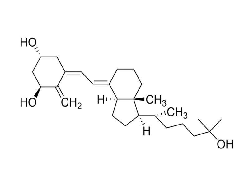
Calcitriol

Vitamin D receptor activator

Calcitriol

Vitamin D receptor activator

From: 373 CAD
Catalog #
(Select a product)
Vitamin D receptor activator
Add to Wish List

Overview

Calcitriol is synthesized from 25-hydroxy Vitamin D3, the principal circulating form of Vitamin D, via hydroxylation in the kidney. The main physiologic effects of calcitriol are to increase the absorption of calcium at the level of the intestinal epithelium, and to increase the mineralization of bone via the direct stimulation of osteoblasts (Portale et al.)

DIFFERENTIATION
· Induces differentiation of human osteoblasts, alone or in combination with TGFβ (Ingram et al.; Kassem et al.; Wergedal et al.).
· Induces differentiation of chicken embryonic chondrocytes (Gerstenfeld et al.; Tsonis).
· Enhances differentiation of human keratinocytes when grown in the presence of high calcium concentrations (Itin et al.).
Cell Type
Chondrocytes, Keratinocytes, Mesenchymal Stem and Progenitor Cells, Osteoblasts
Species
Human, Mouse, Non-Human Primate, Other, Rat
Application
Differentiation
Area of Interest
Stem Cell Biology
CAS Number
32222-06-3
Chemical Formula
C₂₇H₄₄O₃
Purity
≥ 97%
Pathway
Vitamin D
Target
Vitamin D Receptor

Protocols and Documentation

Find supporting information and directions for use in the Product Information Sheet or explore additional protocols below.

Document Type
Product Name
Catalog #
Lot #
Language
Product Name
Calcitriol
Catalog #
72412
Lot #
All
Language
English
Document Type
Safety Data Sheet
Product Name
Calcitriol
Catalog #
72412
Lot #
All
Language
English

Applications

This product is designed for use in the following research area(s) as part of the highlighted workflow stage(s). Explore these workflows to learn more about the other products we offer to support each research area.

Resources and Publications

Educational Materials (1)

Publications (8)

Production and action of transforming growth factor-beta in human osteoblast cultures: dependence on cell differentiation and modulation by calcitriol. Kassem M et al. European journal of clinical investigation 2000 MAY

Abstract

BACKGROUND: Transforming growth factor beta (TGF-beta) plays an important role in skeletal remodelling. However, few studies have examined its effects on cultured human osteoblasts. Our aim is to characterise the biological effects of TGF-beta1 on human osteoblasts and to examine the interaction between TGF-beta1 and calcitriol. DESIGN: In vitro study employing two models of normal human osteoblasts: human bone marrow stromal cells [hMS/(OB)] containing osteoprogenitor cells and trabecular bone osteoblasts (hOB), which are mature osteoblasts. A reverse-transcriptase-polymerase-chain-reaction assay was employed to measure steady state mRNA levels of TGF-beta(s) isoforms and receptors. Effects of short-term treatment of TGF-beta1 on osteoblast proliferation and differentiation markers were assessed. The effect of cotreatment of calcitriol (10-8 M) and TGF-beta1 on osteoblast differentiation was also determined. RESULTS: Both hMS(OB) and hOB cells expressed mRNA transcripts of TGF-beta1, TGF-beta2, TGF-beta 3, TGF-beta type I and type II receptors. TGF-beta 1 stimulated osteoblast proliferation in hMS(OB) and in hOB cultures. In hOB cultures, TGF-beta1 stimulated AP production and cotreatment with calcitriol induced a synergistic increase in AP levels to 250 +/- 61% of calcitriol-treated controls. Effects of TGF-beta1 and calcitriol were less pronounced in hMS(OB) cultures. TGF-beta1 inhibited collagen type I production in hMS(OB) cells and these effects were abolished in presence of calcitriol. In presence of calcitriol, TGF-beta1 increased collagen type I production in hOB cells. In both hOB and hMS(OB) cultures, TGF-beta1 inhibited osteocalcin production. CONCLUSIONS: TGF-beta increases osteoblastic cell proliferation irrespective of the differentiation state. In presence of calcitriol, it initiates osteoblast cell differentiation and matrix formation. As TGF-beta inhibits osteocalcin production, other factors are necessary for inducing terminal differentiation of osteoblasts. The observed effects of TGF-beta on human osteoblasts in vitro may represent important regulatory steps in controlling osteoblast cell proliferation and differentiation in vivo.
Effects of vitamin D metabolites on proliferation and differentiation of cultured human epidermal keratinocytes grown in serum-free or defined culture medium. Itin PH et al. Endocrinology 1994 NOV

Abstract

We examined the effects of 1,25-dihydroxyvitamin D3 [1,25(OH)2D3], 25-hydroxyvitamin D3 (25OHD3), and vitamin D3 on human keratinocyte proliferation and differentiation in a serum-free or defined culture system. Concentrations greater than 10(-8) M 1,25-(OH)2D3 or 10(-7) M 25(OH)2D3 caused marked inhibition of cell growth. Growth inhibition with high doses of 1,25-(OH)2D3 was not stringent, but was mainly exerted in the G1 phase of the cell cycle. Early release from the cell cycle block restored the proliferation of human keratinocytes. The calcium concentration in the medium had no significant effect on the antiproliferative action of 1,25-(OH)2D3, 25OHD3, and vitamin D3. We also show that human keratinocyte proliferation is enhanced at doses of 1,25-(OH)2D3 and 25OH2D3 of 10(-9) M or less. Enhanced proliferation of human keratinocytes with physiological concentrations of 1,25-(OH)2D3 could only be shown in fully defined medium that contained no vitamin D3, related sterols, or bovine pituitary extract. Human keratinocyte differentiation was enhanced with higher doses of 1,25-(OH)2D3 when cells were grown in the presence of high calcium concentrations. These studies demonstrate that the lower, physiological concentrations of vitamin D3 metabolites are capable of stimulating the proliferation of epidermal keratinocytes grown under selected conditions that eliminate confounding or unidentified medium culture factors. Vitamin D3 metabolites are shown to exert mitogenic trophic effects in cultured human epithelial cells similar to their established activities in vivo.
Effects of transforming growth factor beta (TGF beta) and 1,25 dihydroxyvitamin D3 on the function, cytochemistry and morphology of normal human osteoblast-like cells. Ingram RT et al. Differentiation; research in biological diversity 1994 JAN

Abstract

Individually, transforming growth factor beta (TGF beta) and 1,25-dihydroxyvitamin D3 (1,25(OH)2D3) alter the growth and differentiation of normal and transformed osteoblast-like (OB) cells. Although recent evidence suggests interactions between TGF beta and 1,25(OH)2D3 may occur, little is known of the individual or combined effects of these hormones on the expression of the osteoblast phenotype at the cytochemical and biochemical levels in normal human OB (hOB) cells. Primary cultures of hOBs were treated with TGF beta (0.001-10 ng/ml) and 1,25(OH)2D3 (0.1 pM-100 nM) either alone or in combination. TGF beta and 1,25(OH)2D3 stimulated spindle-shaped cells to become stellate in appearance and increased the number of cytoplasmic processes. TGF beta increased 3H-thymidine incorporation and 1,25(OH)2D3 reduced this effect. Conversely, procollagen type-I synthesis and secretion were increased in a dose-dependent manner in the presence of TGF beta but were not significantly affected in the presence of 1,25(OH)2D3. TGF beta and 1,25(OH)2D3 each marginally increased alkaline phosphatase (ALP) activity, but the combination synergistically increased ALP activity in a dose- and time-dependent manner at the cytochemical and biochemical level (three to tenfold over vehicle controls; n = 12). In contrast, TGF beta reduced 1,25(OH)2D3-stimulated osteocalcin secretion. These data suggest that TGF beta stimulates hOB cells to actively produce collagen matrix and proliferate. The combination of TGF beta and 1,25(OH)2D3, however, produces a synergistic increase in ALP activity and maintenance of collagen synthesis. 1,25(OH)2D3 stimulation may induce cells to advance to an endstage where cell proliferation is reduced and osteocalcin expression is promoted. Interactions between TGF beta and 1,25(OH)2D3 may represent important steps in the regulation of osteoblast differentiation and matrix production.