Thank you for your interest in this product.
Please provide us with your contact information and your local representative
will contact you with a customized quote. Where appropriate, they can also assist you with a(n):
Estimated delivery time for your area
Product sample or exclusive offer
In-lab demonstration
By submitting this form, you are providing your consent to STEMCELL Technologies Canada Inc. and its subsidiaries and affiliates (“STEMCELL”) to collect and use your information, and send you newsletters and emails in accordance with our privacy policy. Please contact us with any questions that you may have. You can unsubscribe or change your email preferences at any time.
The GoH3 antibody reacts with CD49f (integrin α6), an ~150 kDa transmembrane glycoprotein that associates non-covalently with CD29 (integrin β1) or CD104 (integrin β4) to form the heterodimeric receptors VLA-6 and α6β4, which bind the extracellular matrix protein laminin. CD49f is a disulfide-linked dimer comprising an ~120 kDa heavy chain and an ~30 kDa membrane-bound light chain. Splice variants exist, which affect the cytoplasmic domain of the protein. CD49f is expressed on the surface of T cells, monocytes, platelets, placental trophoblasts, epithelial cells, and endothelial cells. It is involved in cell adhesion and regulating signaling pathways involved in a variety of processes, including the activation and proliferation of T cells, and the differentiation and maintenance of stem cell pluripotency. CD49f is considered the most important marker for selecting mouse mammary stem and progenitor cells. The GoH3 antibody reacts with an extracellular epitope on CD49f and reportedly blocks integrin α6 function in vivo and binding of integrin α6 to laminin in vitro.
Flow cytometry analysis of human peripheral blood mononuclear cells (PBMCs) labeled with Anti-Mouse CD49f Antibody, Clone GoH3, Alexa Fluor® 488 (filled histogram) or a rat IgG2a, kappa Alexa Fluor® 488 isotype control antibody (solid line histogram).
Figure 2. Data for PE-Conjugated
Flow cytometry analysis of human peripheral blood mononuclear cells (PBMCs) labeled with Anti-Mouse CD49f Antibody, Clone GoH3, PE (filled histogram) or a rat IgG2a, kappa PE isotype control antibody (solid line histogram).
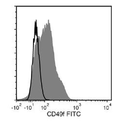
Figure 3. Data for Unconjugated
Flow cytometry analysis of human peripheral blood mononuclear cells (PBMCs) labeled with Anti-Mouse CD49f Antibody, Clone GoH3, followed by a mouse anti-rat IgG2a antibody, FITC (filled histogram), or Rat IgG2a, kappa Isotype Control Antibody, Clone RTK2758 (Catalog #60076), followed by a mouse anti-rat IgG2a antibody, FITC (solid line histogram).
Figure 4. Data for APC-Conjugated
Flow cytometry analysis of human peripheral blood mononuclear cells (PBMCs) labeled with Anti-Mouse CD49f Antibody, Clone GoH3, APC (filled histogram) or Rat IgG2a, kappa Isotype Control Antibody, Clone RTK2758, APC (Catalog #60076AZ) (solid line histogram).
Figure 5. Data for Biotin-Conjugated
Flow cytometry analysis of human peripheral blood mononuclear cells (PBMCs) labeled with Anti-Mouse CD49f Antibody, Clone GoH3, Biotin, followed by streptavidin (SAV) APC (filled histogram), or Rat IgG2a, kappa Isotype Control Antibody, Clone RTK2758, Biotin (Catalog #60076BT), followed by SAV APC (solid line histogram).
Figure 6. Data for FITC-Conjugated
Flow cytometry analysis of human peripheral blood mononuclear cells (PBMCs) labeled with Anti-Mouse CD49f Antibody, Clone GoH3, FITC (filled histogram) or Rat IgG2a, kappa Isotype Control Antibody, Clone RTK2758, FITC (Catalog #60076FI) (solid line histogram).
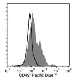
Figure 7. Data for PB-Conjugated
Flow cytometry analysis of human peripheral blood mononuclear cells (PBMCs) labeled with Anti-Mouse CD49f Antibody, Clone GoH3, Pacific Blue™ (filled histogram) or a rat IgG2a, kappa Pacific Blue™ isotype control antibody (solid line histogram).
This product is designed for use in the following research area(s) as part
of the highlighted workflow stage(s). Explore these workflows to learn more about the other products we
offer to support each research area.
Fibroblast Growth Factor Receptor Signaling Is Essential for Normal Mammary Gland Development and Stem Cell Function
Pond AC et al.
Stem cells (Dayton, Ohio) 2013
Abstract
Fibroblast growth factor (FGF) signaling plays an important role in embryonic stem cells and adult tissue homeostasis, but the function of FGFs in mammary gland stem cells is less well defined. Both FGFR1 and FGFR2 are expressed in basal and luminal mammary epithelial cells (MECs), suggesting that together they might play a role in mammary gland development and stem cell dynamics. Previous studies have demonstrated that the deletion of FGFR2 resulted only in transient developmental defects in branching morphogenesis. Using a conditional deletion strategy, we investigated the consequences of FGFR1 deletion alone and then the simultaneous deletion of both FGFR1 and FGFR2 in the mammary epithelium. FGFR1 deletion using a keratin 14 promoter-driven Cre-recombinase resulted in an early, yet transient delay in development. However, no reduction in functional outgrowth potential was observed following limiting dilution transplantation analysis. In contrast, a significant reduction in outgrowth potential was observed upon the deletion of both FGFR1 and FGFR2 in MECs using adenovirus-Cre. Additionally, using a fluorescent reporter mouse model to monitor Cre-mediated recombination, we observed a competitive disadvantage following transplantation of both FGFR1/R2-null MECs, most prominently in the basal epithelial cells. This correlated with the complete loss of the mammary stem cell repopulating population in the FGFR1/R2-attenuated epithelium. FGFR1/R2-null MECs were partially rescued in chimeric outgrowths containing wild-type MECs, suggesting the potential importance of paracrine mechanisms involved in the maintenance of the basal epithelial stem cells. These studies document the requirement for functional FGFR signaling in mammary stem cells during development.
Alexa Fluor and Pacific Blue are trademarks of Life Technologies Corporation. Antibodies conjugated to Alexa Fluor® or Pacific Blue™ are licensed for internal research use only and sale is expressly conditioned on the buyer not using the antibody for manufacturing, performing a service or medical test, or otherwise generating revenue. For use other than research, contact Life Technologies Corporation, 5791 Van Allen Way, Carlsbad, CA 92008 USA or outlicensing@lifetech.com.
Quality Statement:
PRODUCTS ARE FOR RESEARCH USE ONLY AND NOT INTENDED FOR HUMAN OR ANIMAL DIAGNOSTIC OR THERAPEUTIC USES UNLESS OTHERWISE STATED. FOR ADDITIONAL INFORMATION ON QUALITY AT STEMCELL, REFER TO WWW.STEMCELL.COM/COMPLIANCE.