Thank you for your interest in this product.
Please provide us with your contact information and your local representative
will contact you with a customized quote. Where appropriate, they can also assist you with a(n):
Estimated delivery time for your area
Product sample or exclusive offer
In-lab demonstration
By submitting this form, you are providing your consent to STEMCELL Technologies Canada Inc. and its subsidiaries and affiliates (“STEMCELL”) to collect and use your information, and send you newsletters and emails in accordance with our privacy policy. Please contact us with any questions that you may have. You can unsubscribe or change your email preferences at any time.
The 3C7 antibody reacts with murine CD25 (interleukin‐2 receptor α chain; IL‐2Rα), an ~55 kDa type 1 transmembrane glycoprotein expressed on the surface of T and B cell progenitors and activated mature B and T cells. CD25 has been used as a marker to identify CD4+/FoxP3+ regulatory T cells in mice. Mutations in CD25 result in severe immunodeficiency, underscoring the importance of the roles the protein plays in the differentiation, activation and proliferation of lymphocytes, and in the maintenance of self-tolerance. CD25 per se has low affinity for its IL-2 ligand but forms dimers with CD122 (IL-2Rβ) and CD132 (IL-2Rγ) that together associate to form the IL-2R receptor, which binds IL-2 with high affinity. CD25 acts to increase the specificity and affinity of IL-2 binding and is necessary for receptor clustering and signal transduction by the complex. Binding of the 3C7 antibody inhibits binding of IL‐2 to free CD25 and the IL-2R receptor. The epitope is distinct from those recognized by the anti-CD25 antibody clones 7D4 and PC61.
This antibody clone has been verified for purity assessments of cells isolated with EasySep™ kits, including EasySep™ Mouse T Cell Enrichment Kit (Catalog #19751).
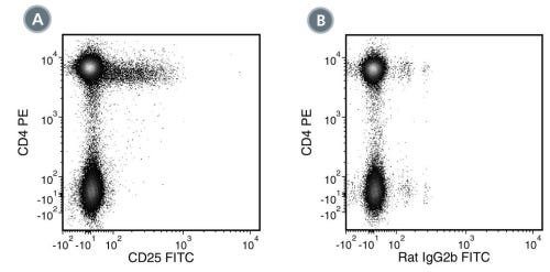
(A) Flow cytometry analysis of C57BL/6 mouse splenocytes (gated on CD3e+ cells) labeled with Anti-Mouse CD25 Antibody, Clone 3C7, followed by a mouse anti-rat IgG2b antibody, FITC and Anti-Mouse CD4, Clone RM4-5, PE (Catalog #60017PE).
(B) Flow cytometry analysis of C57BL/6 mouse splenocytes (gated on CD3e+ cells) labeled with Rat IgG2b, kappa Isotype Control Antibody, Clone RTK4530 (Catalog #60077), followed by a mouse anti-rat IgG2b antibody, FITC and Anti-Mouse CD4, Clone RM4-5, PE.
Figure 2. Data for FITC-Conjugated
(A) Flow cytometry analysis of C57BL/6 mouse splenocytes labeled with Anti-Mouse CD25 Antibody, Clone 3C7, FITC and anti-mouse CD3 APC. (B) Flow cytometry analysis of C57BL/6 mouse splenocytes labeled with a rat IgG2b, kappa FITC isotype control antibody and anti-mouse CD3 APC. (C) Flow cytometry analysis of mouse T cells activated with antibodies against CD3 and CD28. C57BL/6 mouse splenocytes were processed with the EasySep™ Mouse T Cell Enrichment Kit and the enriched T cells were cultured in the absence (Unstimulated) or presence (Stimulated) of Anti-Mouse CD28 and plate-bound Anti-Mouse CD3e for 48 hours, then labeled with Anti-Mouse CD25 Antibody, Clone 3C7, FITC and analyzed for CD25 expression. Upregulation of CD25 following stimulation with CD3 and CD28 is apparent (Stimulated). Labeling of C57BL/6 mouse splenocytes with a rat IgG2b, kappa Alexa Fluor® 488 isotype control antibody is shown (open histogram).
Figure 3. Data for PE-Conjugated
(A) Flow cytometry analysis of C57BL/6 mouse splenocytes labeled with Anti-Mouse CD25 Antibody, Clone 3C7, PE and anti-mouse CD3 APC. (B) Flow cytometry analysis of C57BL/6 mouse splenocytes labeled with a rat IgG2b, kappa PE isotype control antibody and anti-mouse CD3 APC. (C) Flow cytometry analysis of mouse T cells activated with antibodies against CD3 and CD28. C57BL/6 mouse splenocytes were processed with the EasySep™ Mouse T Cell Enrichment Kit (Catalog #19751) and the enriched T cells were cultured in the absence (Unstimulated) or presence (Stimulated) of Anti-Mouse CD28 and plate-bound Anti-Mouse CD3e for 48 hours, then labeled with Anti-Mouse CD25 Antibody, Clone 3C7, PE and analyzed for CD25 expression. Upregulation of CD25 following stimulation with CD3 and CD28 is apparent (Stimulated). Labeling of C57BL/6 mouse splenocytes with a rat IgG2b, kappa Alexa Fluor® 488 isotype control antibody is shown (open histogram).
This product is designed for use in the following research area(s) as part
of the highlighted workflow stage(s). Explore these workflows to learn more about the other products we
offer to support each research area.
Rat monoclonal IgG2b, kappa isotype control antibody
Item added to your cart
Anti-Mouse CD25 Antibody, Clone 3C7
Quality Statement:
PRODUCTS ARE FOR RESEARCH USE ONLY AND NOT INTENDED FOR HUMAN OR ANIMAL DIAGNOSTIC OR THERAPEUTIC USES UNLESS OTHERWISE STATED. FOR ADDITIONAL INFORMATION ON QUALITY AT STEMCELL, REFER TO WWW.STEMCELL.COM/COMPLIANCE.