EasySep™ Human T Cell Isolation Kit

Immunomagnetic negative isolation of untouched human T cells

EasySep™ Human T Cell Isolation Kit

Immunomagnetic negative isolation of untouched human T cells
From: 1,009 USD
Catalog #
(Select a product)
Immunomagnetic negative isolation of untouched human T cells
Add to Wish List

Product Advantages


  • Fast, easy-to-use and column-free

  • Up to 98% purity with high recovery

  • Untouched, viable cells

What's Included

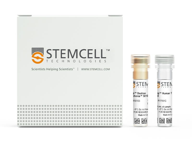
  • EasySep™ Human T Cell Isolation Kit (Catalog #17951)
    • EasySep™ Human T Cell Isolation Cocktail, 1 mL
    • EasySep™ Dextran RapidSpheres™, 1 mL
  • EasySep™ Human T Cell Isolation Kit (Catalog #100-0695)
    • EasySep™ Human T Cell Isolation Cocktail, 1 x 10 mL
    • EasySep™ Dextran RapidSpheres™, 1 x 10 mL
  • RoboSep™ Human T Cell Isolation Kit (Catalog #17951RF)
    • EasySep™ Human T Cell Isolation Cocktail, 1 mL
    • EasySep™ Dextran RapidSpheres™, 1 mL
    • RoboSep™ Buffer (Catalog #20104)
    • RoboSep™ Filter Tips (Catalog #20125)
Products for Your Protocol
To see all required products for your protocol, please consult the Protocols and Documentation.

What Our Scientist Says

Isolating T cells doesn't have to take a long time. We developed this 8-minute T cell isolation kit so you can get to your downstream experiments sooner.

Neil MacDonaldTechnical Scientist
Neil MacDonald, Technical Scientist

Overview

Easily and efficiently isolate highly purified human T cells from fresh or previously frozen human peripheral blood mononuclear cells (PBMCs) or washed leukapheresis samples by immunomagnetic negative selection, with the EasySep™ Human T Cell Isolation Kit. Widely used in published research for more than 20 years, EasySep™ combines the specificity of monoclonal antibodies with the simplicity of a column-free magnetic system.

In this EasySep™ negative selection procedure, unwanted cells are labeled with antibody complexes and magnetic particles. Unwanted cells expressing the following markers are targeted for removal: CD14, CD16, CD19, CD20, CD36, CD56, CD66b, CD123, and GlyA. The magnetically labeled cells are then separated from the untouched desired T cells by using an EasySep™ magnet and simply pouring or pipetting the desired cells into a new tube. Following magnetic cell isolation in as little as 8 minutes, the desired T cells are ready for downstream applications such as flow cytometry, culture, or DNA/RNA extraction.

This product replaces EasySep™ Human T Cell Enrichment Kit (Catalog #19051) for even faster cell isolations.

For large-scale isolation of human T cells from leukapheresis samples, see the large-format (1x10^10 cells) kit (Catalog #100-0695).

Learn more about how immunomagnetic EasySep™ technology works or how to fully automate immunomagnetic cell isolation with RoboSep™. Alternatively, choose ready-to-use, ethically sourced, primary Human Peripheral Blood Pan-T Cells, Frozen isolated with EasySep™ Human T Cell Isolation Kit. Explore additional products optimized for your workflow, including culture media, supplements, antibodies, and more.
Magnet Compatibility
• EasySep™ Magnet (Catalog #18000)
• “The Big Easy” EasySep™ Magnet (Catalog #18001)
• Easy 50 EasySep™ Magnet (Catalog #18002)
• EasyPlate™ EasySep™ Magnet (Catalog 18102)
• EasyEights™ EasySep™ Magnet (Catalog #18103)
• RoboSep™-S (Catalog #21000)
• Easy 250 EasySep™ Magnet (Catalog #100-0821)
Subtype
Cell Isolation Kits
Cell Type
Lymphocytes, T Cells, T Cells, CD3+
Species
Human
Sample Source
Leukapheresis, PBMC
Selection Method
Negative
Application
Cell Isolation
Brand
EasySep, RoboSep
Area of Interest
Chimerism, HLA, Immunology, Cell Therapy Development

Data Figures

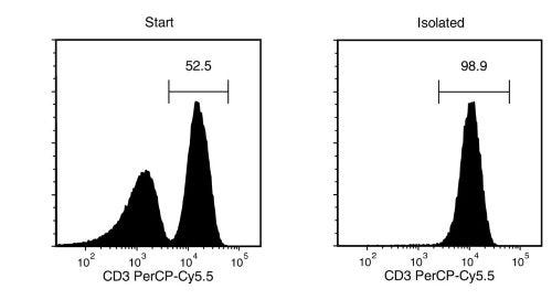
T Cell Separation using EasySep™ Human T Cell Isolation Kit

Figure 1. EasySep™ Human T Cell Isolation Kit

Starting with human peripheral blood mononuclear cells (PBMCs), the T cell content (CD3+) of the isolated fraction is typically 96.7 ± 1.5% (mean ± SD).

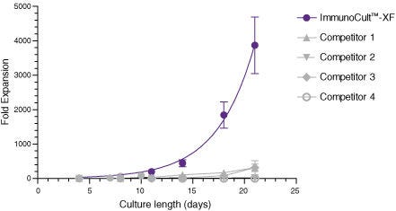
ImmunoCult™-XF T Cell Expansion Medium Supports Faster T Cell Expansion Than Other Serum-Free and Serum-Supplemented Media

Figure 2. ImmunoCult™-XF T Cell Expansion Medium Supports Faster T Cell Expansion Than Other Serum-Free and Serum-Supplemented Media

T cells were isolated from human peripheral blood samples using the EasySep™ Human T Cell Isolation Kit (Catalog #17951), stimulated with ImmunoCult™ Human CD3/CD28/CD2 T Cell Activator (Catalog #10970), and cultured in ImmunoCult™-XF T Cell Expansion Medium supplemented with rhIL-2. T cells were stimulated with ImmunoCult™ Human CD3/CD28/CD2 T Cell Activator on Day 0 and every 7 to 8 days for the duration of the culture. T cells were analyzed on Days 4, 7, 8, 10, 11, 14, 18, and 21 for fold expansion relative to the initial cell seeding density. Compared to all competitor media tested, ImmunoCult™-XF T Cell Expansion Medium showed significantly higher expansion of total T cells. Commercial alternatives 1 to 4 include, in no particular order, X-VIVO™ 15 (Lonza), AIM V® Medium (Life Tech), CellGro® DC Medium (CellGenix), and RPMI 1640 + serum. Each data point represents the mean fold expansion ± S.E.M. at the specified time points (p<0.05 for ImmunoCult™-XF versus all media for Days 8, 11, 14, 18, and 21, tested using two-tailed, paired t-test with unequal variance, n = 6 to 19 donors). The average fold expansion of T cells in ImmunoCult™-XF T Cell Expansion Medium were 15-fold on Day 7, 80-fold on Day 10, 450-fold on Day 14, and 4,000-fold on Day 21.

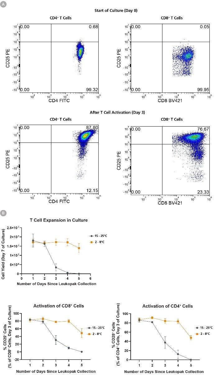
T Cell Expansion and Activation Ability Is Better Preserved When Leukopaks are Stored at Fridge Temperature

Figure 3. T Cell Expansion and Activation Ability Is Better Preserved When Leukopaks are Stored at Fridge Temperature

Using EasySep™ Human T Cell Isolation Kit (Catalog #17951), T cells were isolated from 1 leukopak fraction (Catalog # 70500) of each storage condition daily for 5 days, and 1 x 10⁶ isolated cells were cultured in ImmunoCult™-XF T Cell Expansion Medium (Catalog #10981) supplemented with ImmunoCult™ Human CD3/CD28/CD2 T Cell Activator (Catalog # 10970) and 10 ng/mL IL-2 for 10 days with assessment of CD25 activation marker expression on day 3 of culture. (A) Representative flow cytometry data from leukopaks stored 1 day at fridge temperature (FT), showing that both CD4+ and CD8+ cells are CD25-negative at the start of culture (Day 0), and upregulate CD25 expression by Day 3 of culture. (B) Cellular expansion and corresponding cell yield over 7 days of culture decreases in correlation with storage duration of leukopak fractions. Leukopaks that were stored for 1 - 2 days at either room temperature (RT) or FT had high expansion potential yielding 1.5 - 2 x 10⁷ cells, and this 15 - 20-fold expansion potential is maintained in T cells from leukopaks stored at FT for up to 5 days. In contrast, little or no expansion is observed in T cell cultures from leukopaks stored at RT for 3 or more days, indicating a loss of proliferative capacity. Moreover, T cells show a gradual reduction in their ability to become activated by ImmunoCult™ T Cell activator, as shown by a reduction in Day 3 CD25 expression in gated CD4+ (bottom left) or CD8+ (bottom right) cells over time, and the effect is most pronounced with storage at RT for 3 or more days. All data points represent average ± standard deviation values from leukopak fractions of n = 3 unique donors.

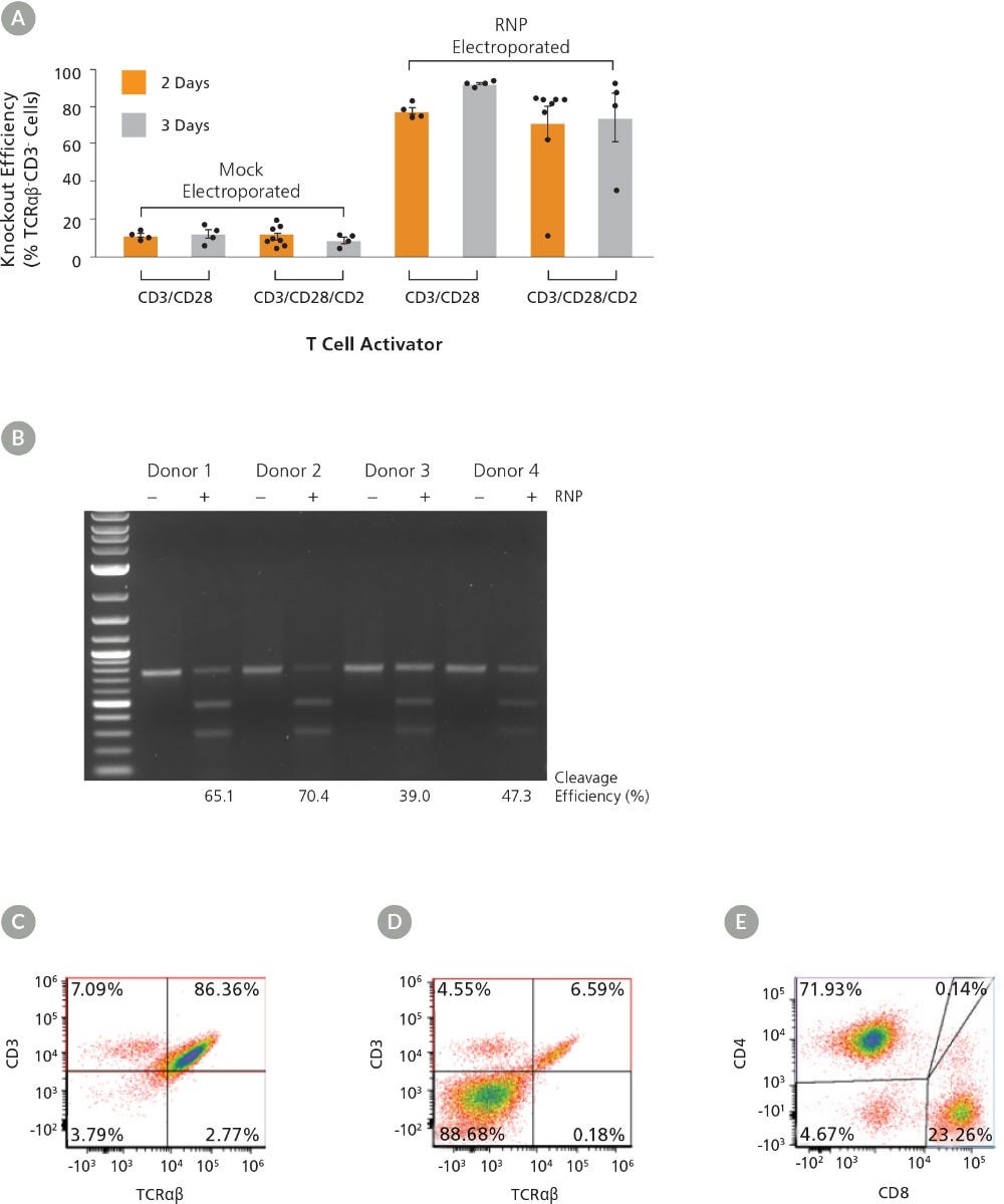
ImmunoCult™ Human T Cell Activators Can Be Used to Optimize Culture Conditions for High-Efficiency TRAC Knockout from Human Primary T Cells

Figure 4. ImmunoCult™ Human T Cell Activators Can Be Used to Optimize Culture Conditions for High-Efficiency TRAC Knockout from Human Primary T Cells

The TRAC locus of human primary T cells was edited with an RNP-based-CRISPR-Cas9 system using multiple T cell activation reagents and dynamics then evaluated to identify a condition with the highest knockout efficiency. (A) TRAC knockout efficiency in human T cells isolated using EasySep™ Human T Cell Isolation Kit (Catalog #17951) and activated with either ImmunoCult™ Human CD3/CD28 or CD3/CD28/CD2 T Cell Activator (Catalog #10971/10970) for 2 or 3 days was assessed by binding the TCRαβ and CD3 receptors with antibodies and performing flow cytometry analysis. Each data point per condition represents an individual donor; n = 4 - 8 donors. Error bars represent standard error of the mean. (B) Genome editing (cleavage) efficiency was assessed at 48 hours post electroporation in human T cells activated with ImmunoCult™ Human CD3/CD28 T Cell Activator for 3 days using the ArciTect™ T7 Endonuclease I Kit (Catalog #76021). Mock electroporated: - RNP; RNP electroporated: + RNP. (C - D) Representative dot plots of TCRαβ and CD3 flow cytometry analysis from (C) mock electroporated and (D) RNP electroporated human T cells activated with ImmunoCult™ Human CD3/CD28 T Cell Activator for 3 days. (E) Representative dot plot of CD4 and CD8 flow cytometry analysis of human T cells activated with ImmunoCult™ Human CD3/CD28 T Cell Activator for 3 days.

Protocols and Documentation

Find supporting information and directions for use in the Product Information Sheet or explore additional protocols below.

Document Type
Product Name
Catalog #
Lot #
Language
Catalog #
17951RF
Lot #
All
Language
English
Catalog #
17951
Lot #
All
Language
English
Catalog #
100-0695
Lot #
All
Language
English
Document Type
Safety Data Sheet 1
Catalog #
17951RF
Lot #
All
Language
English
Document Type
Safety Data Sheet 2
Catalog #
17951RF
Lot #
All
Language
English
Document Type
Safety Data Sheet 3
Catalog #
17951RF
Lot #
All
Language
English
Document Type
Safety Data Sheet 1
Catalog #
17951
Lot #
All
Language
English
Document Type
Safety Data Sheet 2
Catalog #
17951
Lot #
All
Language
English
Document Type
Safety Data Sheet 1
Catalog #
100-0695
Lot #
All
Language
English
Document Type
Safety Data Sheet 2
Catalog #
100-0695
Lot #
All
Language
English

Applications

This product is designed for use in the following research area(s) as part of the highlighted workflow stage(s). Explore these workflows to learn more about the other products we offer to support each research area.

Resources and Publications

Educational Materials (26)

Frequently Asked Questions

Can EasySep™ be used for either positive or negative selection?

Yes. The EasySep™ kits use either a negative selection approach by targeting and removing unwanted cells or a positive selection approach targeting desired cells. Depletion kits are also available for the removal of cells with a specific undesired marker (e.g. GlyA).

How does the separation work?

Magnetic particles are crosslinked to cells using Tetrameric Antibody Complexes (TAC). When placed in the EasySep™ Magnet, labeled cells migrate to the wall of the tube. The unlabeled cells are then poured off into a separate fraction.

Which columns do I use?

The EasySep™ procedure is column-free. That's right - no columns!

How can I analyze the purity of my enriched sample?

The Product Information Sheet provided with each EasySep™ kit contains detailed staining information.

Can EasySep™ separations be automated?

Yes. RoboSep™, the fully automated cell separator, automates all EasySep™ labeling and cell separation steps.

Can EasySep™ be used to isolate rare cells?

Yes. We recommend a cell concentration of 2x10⁸ cells/mL and a minimum working volume of 100 µL. Samples containing 2x10⁷ cells or fewer should be suspended in 100 µL of buffer.

Are the EasySep™ magnetic particles FACS-compatible?

Yes, the EasySep™ particles are flow cytometry-compatible, as they are very uniform in size and about 5000X smaller than other commercially available magnetic beads used with column-free systems.

Can the EasySep™ magnetic particles be removed after enrichment?

No, but due to the small size of these particles, they will not interfere with downstream applications.

Can I alter the separation time in the magnet?

Yes; however, this may impact the kit's performance. The provided EasySep™ protocols have already been optimized to balance purity, recovery and time spent on the isolation.

For positive selection, can I perform more than 3 separations to increase purity?

Yes, the purity of targeted cells will increase with additional rounds of separations; however, cell recovery will decrease.

How does the binding of the EasySep™ magnetic particle affect the cells? is the function of positively selected cells altered by the bound particles?

Hundreds of publications have used cells selected with EasySep™ positive selection kits for functional studies. Our in-house experiments also confirm that selected cells are not functionally altered by the EasySep™ magnetic particles.

If particle binding is a key concern, we offer two options for negative selection. The EasySep™ negative selection kits can isolate untouched cells with comparable purities, while RosetteSep™ can isolate untouched cells directly from whole blood without using particles or magnets.

Publications (84)

Tumor-intrinsic NSUN2 orchestrates immunosuppression in lung adenocarcinoma via the m5C-HDAC8-CCL5 axis. W. Meng et al. Experimental hematology & oncology 2026 Sep

Abstract

BACKGROUND: Insufficient T-cell infiltration is a major barrier to the efficacy of immune checkpoint inhibitors (ICIs) in lung adenocarcinoma (LUAD). We aimed to investigate how the tumor-intrinsic m5C methyltransferase NSUN2 shapes the immune landscape of LUAD. METHODS: Nsun2 conditional knockout mice and syngeneic mice models were employed. Single-cell RNA sequencing (scRNA-seq) and m5C sequencing were performed to elucidate the downstream pathway regulated by NSUN2. LUAD specimens from patients receiving neoadjuvant immunotherapy were used to assess the correlation between NSUN2 expression and immune infiltration. RESULTS: Elevated tumoral NSUN2 expression was correlated with a marked lack of CD8+ T-cell infiltration and immunotherapy resistance. Mechanistically, NSUN2 enhances the translation of histone deacetylase HDAC8 in an m⁵C-YBX1-dependent manner, which in turn directly represses the transcription of the chemoattractant CCL5, thereby impairing CD8+ T-cell recruitment into the tumor. NSUN2 depletion reversed this immunosuppressive axis and converted immunologically "cold" tumors into "hot". Tumor-targeted liposomes loaded with the NSUN2 inhibitor synergized with anti-PD-1 therapy to induce significant tumor regression. CONCLUSIONS: Our findings identify NSUN2 as a critical orchestrator of T-cell exclusion in LUAD. It serves as both a candidate predictive biomarker for ICI failure and a promising druggable target. Targeting NSUN2 offers a potential strategy to overcome immunotherapy resistance and improve clinical outcomes in LUAD patients.
Cancers modulate processing and presentation of p53 neoantigens to evade T cell detection. K. Haratani et al. Immunity 2026 Oct

Abstract

TP53 mutations occur early in malignant transformation as truncal events in tumor evolution and are therefore generally present in all descendant tumor cells, creating an immunological vulnerability. Here, we examined the immunogenicity and antigenicity of p53 neoantigens emerging from these truncal mutations. Comprehensive immunopeptidomics revealed that hotspot mutations in human tumors preferentially localize to p53 regions resistant to antigen processing, thereby avoiding display altogether. Moreover, for neoantigens presentable by HLA-A∗02:01 or HLA-B∗07:02 and structurally divergent from corresponding wild-type p53 peptide-HLA complexes, clinical tumors commonly lacked the relevant presenting HLA allele. Tumor cells further resisted T cell killing through increased activity of the aminopeptidase ERAP1, preventing display of high-affinity HLA-A∗02:01 complexes containing an immunogenic p53I195F-derived 11-mer, or by expressing low-affinity HLA-A∗02:01 complexes containing a p53R175H-derived 9-mer with poor antigenicity despite high-quality human TCRs. These findings define mechanisms by which tumors restrict targetable truncal neoantigen display and suggest immunopeptidome shift strategies to circumvent immune escape.
Myeloperoxidase promotes a tumorigenic microenvironment in non-small cell lung cancer. P. Valadez-Cosmes et al. Redox biology 2026 Nov

Abstract

Myeloperoxidase (MPO) is a heme peroxidase that is mainly expressed and secreted by neutrophils. MPO's role in inflammatory diseases has been highlighted in recent years, but its role in tumor development remains unclear. Therefore, we investigated the role of MPO in non-small cell lung cancer (NSCLC). In silico analysis revealed a survival benefit in patients with NSCLC and low MPO expression. Furthermore, a syngeneic tumor model using MPO knockout (KO) mice revealed that mice lacking MPO had lower tumor growth than controls. The reduction in tumor size was accompanied by an increase in lymphoid populations, including natural killer cells and CD8+ T cells, suggesting a shift to a more anti-tumorigenic immune environment in MPO-KO mouse tumors. The T cell induced interferon-gamma (IFN-γ) expression was increased in MPO-KO tumors, indicating increased tumoricidal activity. CD8 depletion abolished the previously observed reduction in tumor size in MPO-KO mice, indicating that CD8+ T cells play an important role. In vitro, T cells treated with MPO showed reduced proliferation and IFN-γ expression. Furthermore, MPO associated directly with T cells. Interestingly, MPO+ lymphocytes, including CD8+ T cells, were found in tumor samples from patients with NSCLC. Our findings suggest that MPO plays an immunosuppressive role in NSCLC.