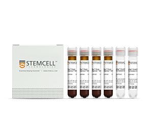
EasySep™ Mouse NK Cell Isolation Kit

25-Minute cell isolation kit using immunomagnetic negative selection
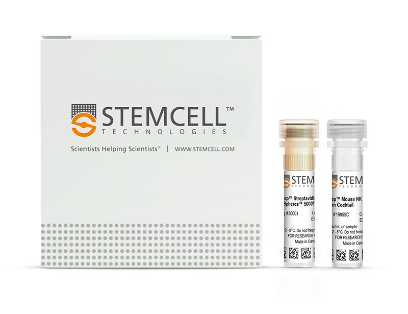
New look, same high quality and support! You may notice that your instrument or reagent packaging looks slightly different from images displayed on the website, or from previous orders. We are updating our look but rest assured, the products themselves and how you should use them have not changed. Learn more

EasySep™ Mouse NK Cell Isolation Kit

25-Minute cell isolation kit using immunomagnetic negative selection

From: 785 USD
Catalog #
(Select a product)
25-Minute cell isolation kit using immunomagnetic negative selection
Add to Wish List

Product Advantages


  • Fast and easy-to-use

  • No columns required

  • Untouched, viable cells

What's Included

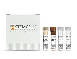
  • EasySep™ Mouse NK Cell Isolation Kit (Catalog #19855)
    • EasySep™ Mouse NK Cell Isolation Cocktail, 0.5 mL
    • EasySep™ Streptavidin RapidSpheres™ 50001, 1 mL
  • RoboSep™ Mouse NK Cell Isolation Kit (Catalog #19855RF)
    • EasySep™ Mouse NK Cell Isolation Cocktail, 0.5 mL
    • EasySep™ Streptavidin RapidSpheres™ 50001, 1 mL
    • RoboSep™ Buffer (Catalog #20104)
    • RoboSep™ Filter Tips (Catalog #20125)

Overview

The EasySep™ Mouse NK Cell Isolation Kit targets non-NK cells by labeling unwanted cells with biotinylated antibodies and EasySep™ Streptavidin RapidSpheres™, and separates cells without columns using an EasySep™ magnet. Desired cells are simply poured off into a new tube. Isolated cells are immediately available for downstream applications such as flow cytometry, culture or cell-based assays.

This product replaces the EasySep™ Mouse NK Cell Enrichment Kit (Catalog #19755) for even faster cell isolations.
Magnet Compatibility
• EasySep™ Magnet (Catalog #18000)
• “The Big Easy” EasySep™ Magnet (Catalog #18001)
• EasyEights™ EasySep™ Magnet (Catalog #18103)
• RoboSep™-S (Catalog #21000)
Subtype
Cell Isolation Kits
Cell Type
NK Cells
Species
Mouse
Sample Source
Spleen
Selection Method
Negative
Application
Cell Isolation
Brand
EasySep, RoboSep
Area of Interest
Immunology

Data Figures

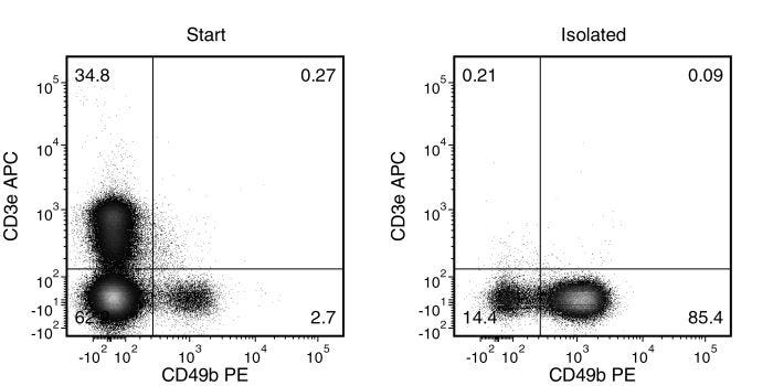
Typical EasySep™ Mouse NK Cell Isolation Profile

Figure 1. Typical EasySep™ Mouse NK Cell Isolation Profile

Starting with mouse splenocytes, the NK cell (CD3-CD49b+) content of the isolated fraction typically ranges from 67 - 89%.

Protocols and Documentation

Find supporting information and directions for use in the Product Information Sheet or explore additional protocols below.

Document Type
Product Name
Catalog #
Lot #
Language
Catalog #
19855RF
Lot #
1000100903 and lower
Language
English
Catalog #
19855RF
Lot #
1000100904 and higher
Language
English
Catalog #
19855
Lot #
1000100903 and lower
Language
English
Catalog #
19855
Lot #
1000100904 and higher
Language
English
Document Type
Safety Data Sheet 1
Catalog #
19855RF
Lot #
All
Language
English
Document Type
Safety Data Sheet 2
Catalog #
19855RF
Lot #
All
Language
English
Document Type
Safety Data Sheet 3
Catalog #
19855RF
Lot #
All
Language
English
Document Type
Safety Data Sheet 1
Catalog #
19855
Lot #
All
Language
English
Document Type
Safety Data Sheet 2
Catalog #
19855
Lot #
All
Language
English

Applications

This product is designed for use in the following research area(s) as part of the highlighted workflow stage(s). Explore these workflows to learn more about the other products we offer to support each research area.

Resources and Publications

Frequently Asked Questions

Can EasySep™ be used for either positive or negative selection?

Yes. The EasySep™ kits use either a negative selection approach by targeting and removing unwanted cells or a positive selection approach targeting desired cells. Depletion kits are also available for the removal of cells with a specific undesired marker (e.g. GlyA).

How does the separation work?

Magnetic particles are crosslinked to cells using Tetrameric Antibody Complexes (TAC). When placed in the EasySep™ Magnet, labeled cells migrate to the wall of the tube. The unlabeled cells are then poured off into a separate fraction.

Which columns do I use?

The EasySep™ procedure is column-free. That's right - no columns!

How can I analyze the purity of my enriched sample?

The Product Information Sheet provided with each EasySep™ kit contains detailed staining information.

Can EasySep™ separations be automated?

Yes. RoboSep™, the fully automated cell separator, automates all EasySep™ labeling and cell separation steps.

Can EasySep™ be used to isolate rare cells?

Yes. We recommend a cell concentration of 2x10⁸ cells/mL and a minimum working volume of 100 µL. Samples containing 2x10⁷ cells or fewer should be suspended in 100 µL of buffer.

Are the EasySep™ magnetic particles FACS-compatible?

Yes, the EasySep™ particles are flow cytometry-compatible, as they are very uniform in size and about 5000X smaller than other commercially available magnetic beads used with column-free systems.

Can the EasySep™ magnetic particles be removed after enrichment?

No, but due to the small size of these particles, they will not interfere with downstream applications.

Can I alter the separation time in the magnet?

Yes; however, this may impact the kit's performance. The provided EasySep™ protocols have already been optimized to balance purity, recovery and time spent on the isolation.

For positive selection, can I perform more than 3 separations to increase purity?

Yes, the purity of targeted cells will increase with additional rounds of separations; however, cell recovery will decrease.

How does the binding of the EasySep™ magnetic particle affect the cells? is the function of positively selected cells altered by the bound particles?

Hundreds of publications have used cells selected with EasySep™ positive selection kits for functional studies. Our in-house experiments also confirm that selected cells are not functionally altered by the EasySep™ magnetic particles.

If particle binding is a key concern, we offer two options for negative selection. The EasySep™ negative selection kits can isolate untouched cells with comparable purities, while RosetteSep™ can isolate untouched cells directly from whole blood without using particles or magnets.

Publications (8)

CRISPR-Cas9 Ribonucleoprotein-Mediated Genomic Editing in Mature Primary Innate Immune Cells. L. Riggan et al. Cell reports 2020 may

Abstract

CRISPR genome engineering has become a powerful tool to functionally investigate the complex mechanisms of immune system regulation. While decades of work have aimed to genetically reprogram innate immunity, the utility of current approaches is restricted by poor knockout efficiencies or limited specificity for mature cell lineages in vivo. Here, we describe an optimized strategy for non-viral CRISPR-Cas9 ribonucleoprotein (cRNP) genomic editing of mature primary mouse innate lymphocyte cells (ILCs) and myeloid lineage cells that results in an almost complete loss of single or double target gene expression from a single electroporation. Furthermore, we describe in vivo adoptive transfer mouse models that can be utilized to screen for gene function during viral infection using cRNP-edited naive natural killer (NK) cells and bone-marrow-derived conventional dendritic cell precursors (cDCPs). This resource will enhance target gene discovery and offer a specific and simplified approach to gene editing in the mouse innate immune system.
NK Cells Are Critical for Optimal Immunity to Experimental Trypanosoma congolense Infection. C. Onyilagha et al. Journal of immunology (Baltimore, Md. : 1950) 2019 jun

Abstract

NK cells are key innate immune cells that play critical roles in host defense. Although NK cells have been shown to regulate immunity to some infectious diseases, their role in immunity to Trypanosoma congolense has not been investigated. NK cells are vital sources of IFN-gamma and TNF-alpha; two key cytokines that are known to play important roles in resistance to African trypanosomes. In this article, we show that infection with T. congolense leads to increased levels of activated and functional NK cells in multiple tissue compartments. Systemic depletion of NK cells with anti-NK1.1 mAb led to increased parasitemia, which was accompanied by significant reduction in IFN-gamma production by immune cells in the spleens and liver of infected mice. Strikingly, infected NFIL3-/- mice (which genetically lack NK cell development and function) on the normally resistant background were highly susceptible to T. congolense infection. These mice developed fulminating and uncontrolled parasitemia and died significantly earlier (13 ± 1 d) than their wild-type control mice (106 ± 26 d). The enhanced susceptibility of NFIL3-/- mice to infection was accompanied by significantly impaired cytokine (IFN-gamma and TNF-alpha) response by CD3+ T cells in the spleens and liver. Adoptive transfer of NK cells into NFIL3-/- mice before infection rescued them from acute death in a perforin-dependent manner. Collectively, these studies show that NK cells are critical for optimal resistance to T. congolense, and its deficiency leads to enhanced susceptibility in infected mice.
Endosomolytic polymersomes increase the activity of cyclic dinucleotide STING agonists to enhance cancer immunotherapy. D. Shae et al. Nature nanotechnology 2019 jan

Abstract

Cyclic dinucleotide (CDN) agonists of stimulator of interferon genes (STING) are a promising class of immunotherapeutics that activate innate immunity to increase tumour immunogenicity. However, the efficacy of CDNs is limited by drug delivery barriers, including poor cellular targeting, rapid clearance and inefficient transport to the cytosol where STING is localized. Here, we describe STING-activating nanoparticles (STING-NPs)-rationally designed polymersomes for enhanced cytosolic delivery of the endogenous CDN ligand for STING, 2'3' cyclic guanosine monophosphate-adenosine monophosphate (cGAMP). STING-NPs increase the biological potency of cGAMP, enhance STING signalling in the tumour microenvironment and sentinel lymph node, and convert immunosuppressive tumours to immunogenic, tumoricidal microenvironments. This leads to enhanced therapeutic efficacy of cGAMP, inhibition of tumour growth, increased rates of long-term survival, improved response to immune checkpoint blockade and induction of immunological memory that protects against tumour rechallenge. We validate STING-NPs in freshly isolated human melanoma tissue, highlighting their potential to improve clinical outcomes of immunotherapy.
New look, same high quality and support! You may notice that your instrument or reagent packaging looks slightly different from images displayed on the website, or from previous orders. We are updating our look but rest assured, the products themselves and how you should use them have not changed. Learn more