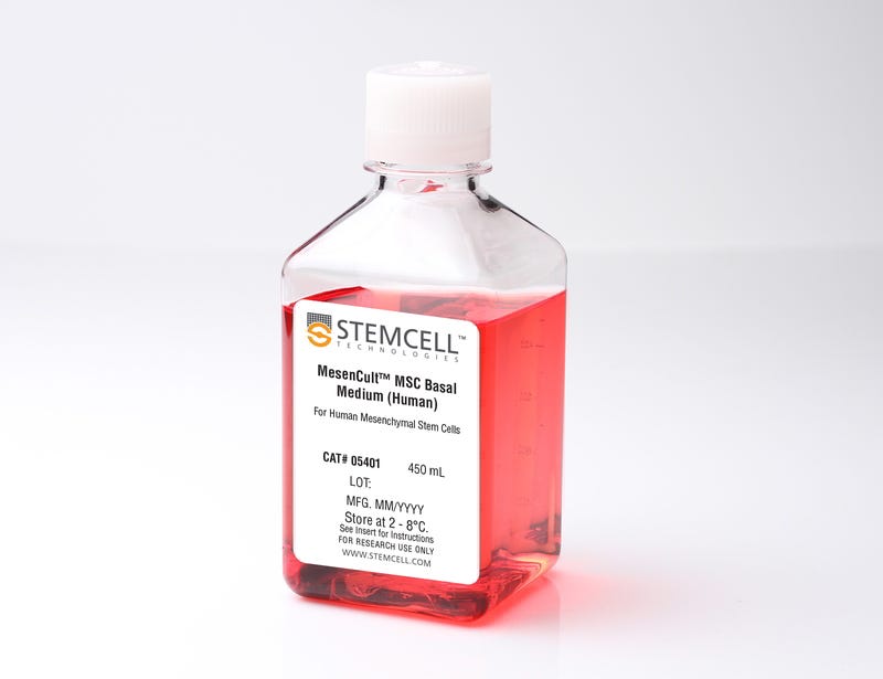
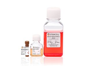
MesenCult™ MSC Basal Medium (Human)

Basal medium for human mesenchymal stem cells

MesenCult™ MSC Basal Medium (Human)

Basal medium for human mesenchymal stem cells

From: 68 USD
Catalog #
(Select a product)
Basal medium for human mesenchymal stem cells
Add to Wish List
Products for Your Protocol
To see all required products for your protocol, please consult the Protocols and Documentation.

Overview

MesenCult™ MSC Basal Medium (Human) is a standardized basal medium designed to be supplemented with MesenCult™ Mesenchymal Stem Cell Stimulatory Supplement (Human; Catalog #05402) for the in vitro culture of human mesenchymal stem cells (MSCs). MesenCult™ MSC Basal Medium is a component of MesenCult™ Proliferation Kit (Human; Catalog #05411), and is also available separately.
Subtype
Basal Media
Cell Type
Mesenchymal Stem and Progenitor Cells
Species
Human
Application
Cell Culture, Colony Assay, Expansion
Brand
MesenCult
Area of Interest
Stem Cell Biology

Protocols and Documentation

Find supporting information and directions for use in the Product Information Sheet or explore additional protocols below.

Document Type
Product Name
Catalog #
Lot #
Language
Catalog #
05401
Lot #
19A99097 or lower
Language
English
Catalog #
05401
Lot #
All
Language
English
Document Type
Safety Data Sheet
Catalog #
05401
Lot #
All
Language
English

Applications

This product is designed for use in the following research area(s) as part of the highlighted workflow stage(s). Explore these workflows to learn more about the other products we offer to support each research area.

Resources and Publications

Publications (48)

FAK Deficiency in Bone Marrow Stromal Cells Alters Their Homeostasis and Drives Abnormal Proliferation and Differentiation of Haematopoietic Stem Cells. Y. Wu et al. Cells 2020 mar

Abstract

Emerging evidence indicates that in myelodysplastic syndromes (MDS), the bone marrow (BM) microenvironment may also contribute to the ineffective, malignant haematopoiesis in addition to the intrinsic abnormalities of haematopoietic stem precursor cells (HSPCs). The BM microenvironment influences malignant haematopoiesis through indirect mechanisms, but the processes by which the BM microenvironment directly contributes to MDS initiation and progression have not yet been elucidated. Our previous data showed that BM-derived stromal cells (BMSCs) from MDS patients have an abnormal expression of focal adhesion kinase (FAK). In this study, we characterise the morpho-phenotypic features and the functional alterations of BMSCs from MDS patients and in FAK knock-downed HS-5 cells. The decreased expression of FAK or its phosphorylated form in BMSCs from low-risk (LR) MDS directly correlates with BMSCs' functional deficiency and is associated with a reduced level of haemoglobin. The downregulation of FAK in HS-5 cells alters their morphology, proliferation, and differentiation capabilities and impairs the expression of several adhesion molecules. In addition, we examine the CD34+ healthy donor (HD)-derived HSPCs' properties when co-cultured with FAK-deficient BMSCs. Both abnormal proliferation and the impaired erythroid differentiation capacity of HD-HSPCs were observed. Together, these results demonstrate that stromal adhesion mechanisms mediated by FAK are crucial for regulating HSPCs' homeostasis.
Icariin protects rabbit BMSCs against OGD-induced apoptosis by inhibiting ERs-mediated autophagy via MAPK signaling pathway. D. Liu et al. Life sciences 2020 jul

Abstract

Stem cell therapy is widely employed in treating osteoarthritis (OA), and bone marrow-derived mesenchymal stem cells (BMSCs) has gradually become the most attractive new method for treating OA due to the benefit for cartilage tissue repair. However, the apoptosis in the neural stem cell transplantation severely decreases repairing efficacy. Icariin has been reported to exert multiple effects on BMSCs, including its proliferation, osteogenic, and chondrogenic differentiation. However, its effects on the injury induced by oxygen, glucose and serum deprivation (OGD) remains unknown. We prospectively investigated the role of ICA on rabbit BMSCs under conditions of OGD. Firstly, BMSCs were cultured under conditions of OGD, ICA relieved OGD-induced cell damage by promoting cell proliferation and suppressing apoptosis. Secondly, Markers of endoplasmic reticulum stress (ERs), ER stress IRE-1 pathway, and autophagy were both inhibited by ICA via inhibition of phosphor-extracellular regulated protein kinases (p-ERKs), p-P38, p-c-Jun N-terminal kinase (p-JNK) or si-MAPK. Finally, decrease of ERs marker levels enhanced protective effect of ICA against OGD-induced injury by limiting apoptosis and autophagy. Moreover, an autophagy inhibitor (3-methyladenine: 3-MA) contributed to a synergistic effect in conjunction with ICA, in promoting cell proliferation, suggesting that ICA exerts anti-ERs and anti-autophagy effects in OGD-treated BMSCs. Therefore, ICA protected rabbit BMSCs from OGD-induced apoptosis through inhibitory regulation of ERs-mediated autophagy related to the MAPK signaling pathway, which provided insights for a potential therapeutic strategy in OA.
A Unique Nonsaccharide Mimetic of Heparin Hexasaccharide Inhibits Colon Cancer Stem Cells via p38 MAP Kinase Activation. R. S. Boothello et al. Molecular cancer therapeutics 2019

Abstract

Targeting of cancer stem cells (CSC) is expected to be a paradigm-shifting approach for the treatment of cancers. Cell surface proteoglycans bearing sulfated glycosaminoglycan (GAG) chains are known to play a critical role in the regulation of stem cell fate. Here, we show for the first time that G2.2, a sulfated nonsaccharide GAG mimetic (NSGM) of heparin hexasaccharide, selectively inhibits colonic CSCs in vivo G2.2-reduced CSCs (CD133+/CXCR4+, Dual hi) induced HT-29 and HCT 116 colon xenografts' growth in a dose-dependent fashion. G2.2 also significantly delayed the growth of colon xenograft further enriched in CSCs following oxaliplatin and 5-fluorouracil treatment compared with vehicle-treated xenograft controls. In fact, G2.2 robustly inhibited CSCs' abundance (measured by levels of CSC markers, e.g., CD133, DCMLK1, LGR5, and LRIG1) and self-renewal (quaternary spheroids) in colon cancer xenografts. Intriguingly, G2.2 selectively induced apoptosis in the Dual hi CSCs in vivo eluding to its CSC targeting effects. More importantly, G2.2 displayed none to minimal toxicity as observed through morphologic and biochemical studies of vital organ functions, blood coagulation profile, and ex vivo analyses of normal intestinal (and bone marrow) progenitor cell growth. Through extensive in vitro, in vivo, and ex vivo mechanistic studies, we showed that G2.2's inhibition of CSC self-renewal was mediated through activation of p38$\alpha$, uncovering important signaling that can be targeted to deplete CSCs selectively while minimizing host toxicity. Hence, G2.2 represents a first-in-class (NSGM) anticancer agent to reduce colorectal CSCs.