TeSR™-E7™ Medium for Reprogramming (2-Component)

Feeder-free and animal component-free reprogramming medium for human iPS cell induction

TeSR™-E7™ Medium for Reprogramming (2-Component)

Feeder-free and animal component-free reprogramming medium for human iPS cell induction

From: 312 USD
Catalog #
(Select a product)
Feeder-free and animal component-free reprogramming medium for human iPS cell induction
Add to Wish List

Product Advantages


  • Pre-screened components ensure high quality iPS cell colony morphology for easy identification and improved manual selection

  • Reduced differentiation and fibroblast growth enables rapid establishment of homogeneous iPS cell cultures

  • Feeder-free, defined formulation facilitates reproducibly efficient human iPS cell generation

What's Included

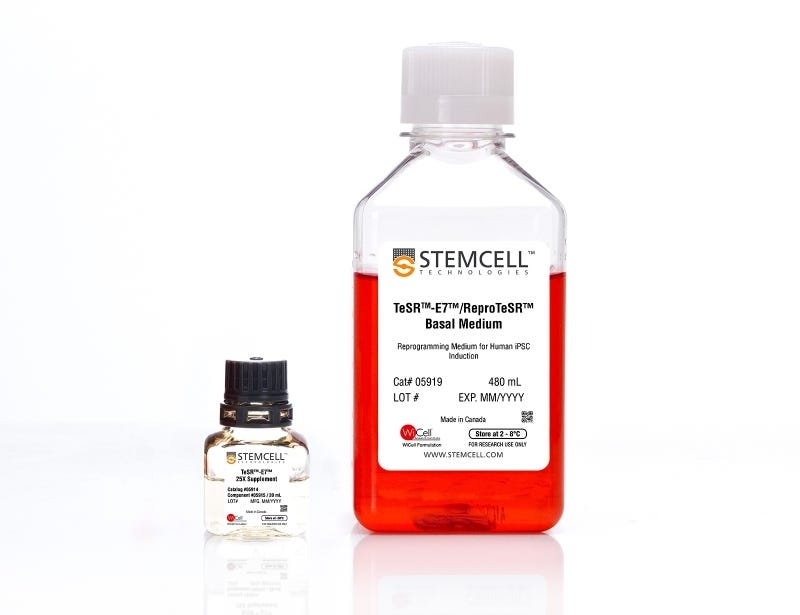
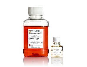
TeSR™-E7™/ ReproTeSR™ Basal Medium, 480 mL
TeSR™-E7™ 25X Supplement, 20 mL
 

Overview

TeSR™-E7™ (2-Component) is an animal component-free and defined reprogramming culture medium optimized for the generation of human iPS cells without the use of feeders. It is based on the E7 formulation published by the laboratory of Dr. James Thomson (University of Wisconsin-Madison).
Subtype
Specialized Media
Cell Type
Pluripotent Stem Cells
Species
Human
Application
Cell Culture, Reprogramming
Brand
TeSR
Area of Interest
Stem Cell Biology
Formulation Category
Animal Component-Free, Serum-Free, Xeno-Free

Data Figures

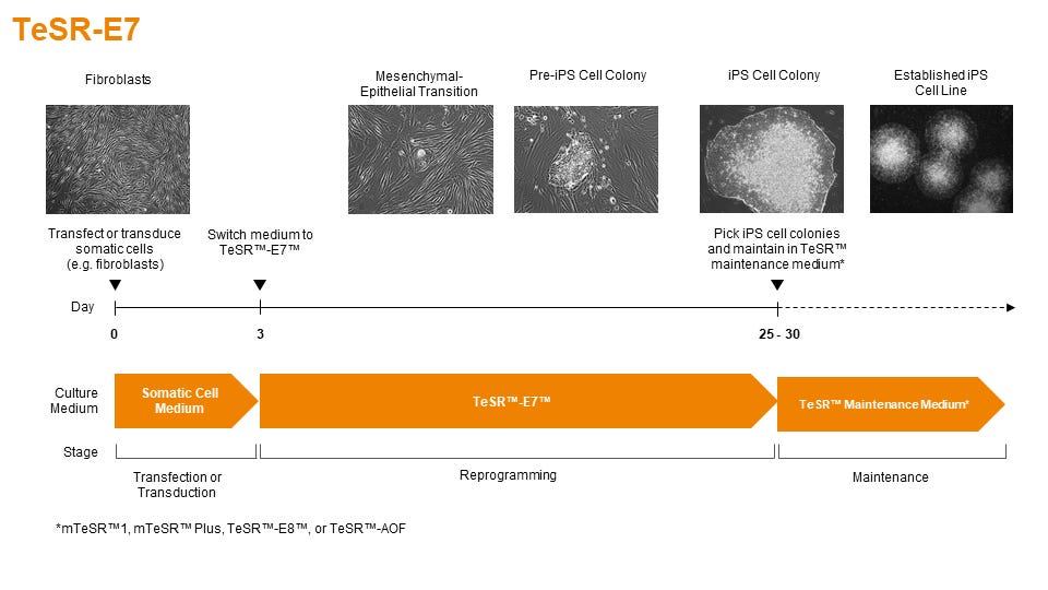
Figure 1. Schematic of Reprogramming Timeline

TeSR™-E7™ can be used during the entire induction phase of reprogramming (day 3 to 25+). Following reprogramming, iPS cell colonies can be isolated and propogated in feeder-free maintenance systems (eg. mTeSR™1 or TeSR™-E8™ media on Corning® Matrigel® or Vitronectin XF™ matrices).

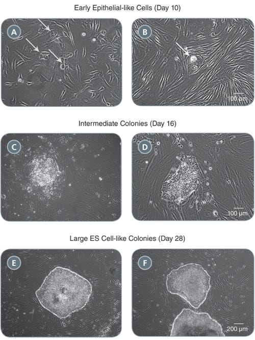
Figure 2. Morphology of Representative iPS Cell Colonies Arising During the Induction Period in TeSR™-E7™

(A-B) Small clusters of colonies with an epithelial-like morphology will appear by one to two weeks following induction (see arrows). (C-D) These clusters expand into pre-iPS cell colonies by two to three weeks. (E-F) Larger ES cell-like colonies are clearly identifiable by three to four weeks. Representative colonies from adult human fibroblasts reprogrammed with episomal vectors containing OCT-4, SOX2, KLF-4, and L-MYC are shown.

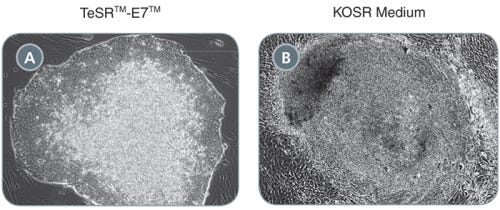
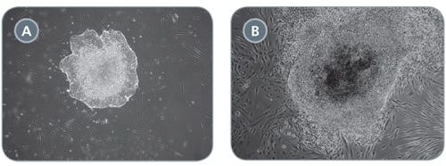
Figure 3. Comparison of Primary iPS Cell Colonies Derived Using TeSR™-E7™ and KOSR-Based Medium

(A) TeSR™-E7™ generates colonies with defined borders and less overgrowth of background fibroblasts compared to (B) KOSR-based iPS cell induction medium. Representative colonies from adult human fibroblasts reprogrammed with episomal vectors containing OCT-4, SOX2, KLF-4, and L-MYC are shown.

Figure 4. Comparison of Primary iPS Cell Colonies Derived Using TeSR™-E7™ with Qualified vs Unqualified bFGF

(A) TeSR™-E7™ yields easily recognizable iPS cell colonies with defined borders. (B) Unqualified components can result in colonies that have poorly defined edges and higher levels of differentiation. Representative colonies from adult human fibroblasts reprogrammed with episomal vectors containing OCT-4, SOX2, KLF-4, and L-MYC are shown.

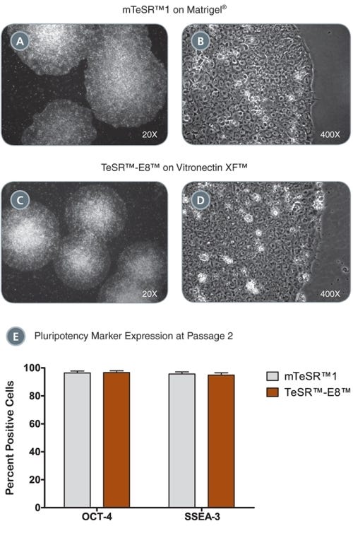
Figure 5. iPS Colonies Expanded in mTeSR™ or TeSR™-E8™

(A - D) iPS cell colonies generated in TeSR™-E7™ and expanded in either mTeSR™1 on Corning® Matrigel® (A-B) or TeSR™-E8™ on Vitronectin XF™ (C, D) exhibit classic ES cell morphology with dense colony centers, defined borders, prominent nucleoli and high nuclear-to-cytoplasmic ratios. (E) iPS cells express high levels of pluripotency markers after just two passages in either mTeSR™1 or TeSR™-E8™ as demonstrated by OCT-4 and SSEA-3 flow cytometry analysis. Data are expressed as mean ± SEM, n = 4.

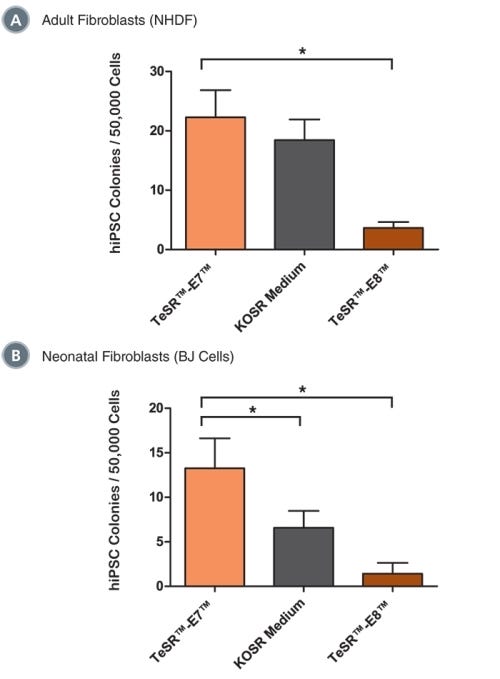
Figure 6. TeSR™-E7™ Supports Reprogramming of Human Cell Types Including Adult Dermal Fibroblasts and Neonatal Fibroblasts

Reprogramming of (A) adult normal human dermal fibroblasts (NHDF, 33 year-old female) and (B) neonatal foreskin fibroblasts (BJ cells) with episomal reprogramming vectors are shown. TeSR™-E7™ demonstrated similar (in NHDF) or greater (in BJ cells) reprogramming efficiencies compared to KOSR-based iPS cell induction medium. TeSR™-E7™ demonstrated higher reprogramming efficiencies compared to TeSR™-E8™. Data are expressed as mean ± SEM, n ≥ 6, * p ≤ 0.05.

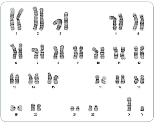
Figure 7. iPS Cells Derived in TeSR™-E7™ Display Normal Karyotype

iPS cell lines were generated in TeSR™-E7™ medium, maintained in mTeSR™1 or TeSR™-E8™ media for a minimum of 5 passages and karyotyped by G-banding karyotype analysis. Three iPS cell lines were analyzed and all demonstrated a normal karyotype; a representative karyogram is shown.

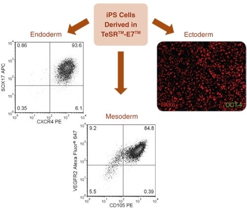
Figure 8. Directed Differentiation of iPS Cells to All Three Germ Layers

TeSR™-E7™-derived iPS cells were differentiated into all three germ layers. Endoderm specification was achieved using the STEMdiff™ Definitive Endoderm Kit, results demonstrated 93.6% SOX17 + CXCR4 + cells. Mesoderm specification was demonstrated using a STEMdiff™ APEL™ medium-based endothelial differentiation protocol, results demonstrated &ht;99% CD31 + cells (data not shown) and 84.8% VEGFR2 + CD105 + cells. Ectoderm specification was demonstrated using STEMdiff™ Neural Induction Medium, immunocytochemistry shows high levels of PAX6 staining with no detectable OCT-4 staining by day 9 of neural induction.

Protocols and Documentation

Find supporting information and directions for use in the Product Information Sheet or explore additional protocols below.

Document Type
Product Name
Catalog #
Lot #
Language
Catalog #
05914
Lot #
All
Language
English
Document Type
Safety Data Sheet 1
Catalog #
05914
Lot #
All
Language
English
Document Type
Safety Data Sheet 2
Catalog #
05914
Lot #
All
Language
English

Applications

This product is designed for use in the following research area(s) as part of the highlighted workflow stage(s). Explore these workflows to learn more about the other products we offer to support each research area.