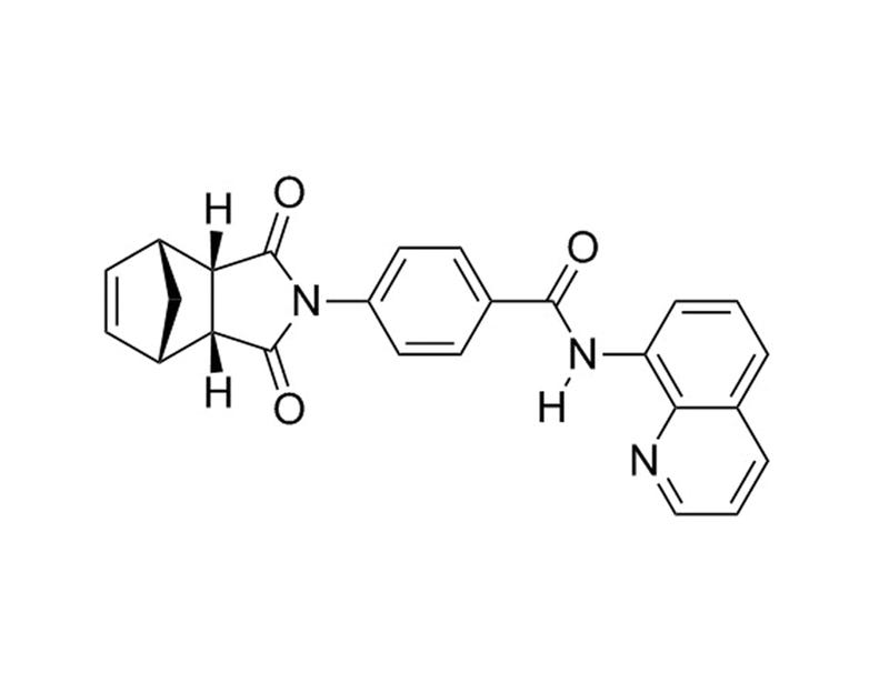
IWR-1-endo

WNT pathway inhibitor; AXIN2 stabilizer

IWR-1-endo

WNT pathway inhibitor; AXIN2 stabilizer
From: 125 USD
Catalog #
(Select a product)
WNT pathway inhibitor; AXIN2 stabilizer
Add to Wish List

Overview

IWR-1-endo potently inhibits WNT signaling by blocking a cell-based WNT/β-catenin pathway reporter response with an IC₅₀ value of 180 nM (Chen et al.). It inhibits WNT-induced accumulation of β-catenin, through stabilization of the destruction complex member AXIN2 (Chen et al.).

MAINTENANCE AND SELF-RENEWAL
· Promotes self-renewal and maintains pluripotency of human embryonic stem cells and mouse Epi-stem cells when used in combination with CHIR99021 (Kim et al.).

DIFFERENTIATION
· Promotes differentiation of cardiomyocytes from human pluripotent stem cells (PSCs) that have been induced to mesoderm by addition of Activin A and/or BMP4 (Ren et al.; Willems et al.)
· Induces the differentiation of human PSC-derived alveolar epithelial type II (AETII) to AETI cells (Ghaedi et al.).
Cell Type
Airway Cells, Cardiomyocytes, PSC-Derived, Pluripotent Stem Cells
Species
Human, Mouse, Non-Human Primate, Other, Rat
Application
Differentiation, Expansion, Maintenance
Area of Interest
Epithelial Cell Biology, Stem Cell Biology
CAS Number
1127442-82-3
Chemical Formula
C₂₅H₁₉N₃O₃
Purity
≥ 98%
Pathway
WNT
Target
Axin

Protocols and Documentation

Find supporting information and directions for use in the Product Information Sheet or explore additional protocols below.

Document Type
Product Name
Catalog #
Lot #
Language
Product Name
IWR-1-endo
Catalog #
72564, 72562
Lot #
All
Language
English
Document Type
Safety Data Sheet
Product Name
IWR-1-endo
Catalog #
72564, 72562
Lot #
All
Language
English

Applications

This product is designed for use in the following research area(s) as part of the highlighted workflow stage(s). Explore these workflows to learn more about the other products we offer to support each research area.

Resources and Publications

Publications (11)

Structural and functional benchmarking of monolayer- and bioreactor-generated hiPSC-derived cardiomyocytes. Y. Jang et al. APL bioengineering 2026 Sep

Abstract

Transitioning from animal to human cell sources represents a critical milestone in cardiac tissue engineering and biomedical research. Neonatal rat ventricular myocytes (NRVMs) have long served as the functional benchmark for engineered cardiac tissues; however, their rodent origin limits clinical relevance. Human-induced pluripotent stem cell-derived cardiomyocytes (hiPSC-CMs) offer a renewable, species-specific alternative but remain restricted by immature structure and function, small-scale yield, and high batch variability by conventional two-dimensional monolayer (2D-Mono) differentiation. Here, we systematically evaluated hiPSC-CMs generated by 2D-Mono and three-dimensional embryoid-body (3D-EB) differentiation using identical 15-day Wnt-modulated protocols without additional maturation steps. The 3D-EB method yielded 181 × 106 cells per 100 ml, approximately 2.7-fold higher than the 2D-Mono, while maintaining >80% cTnT+ purity and reduced batch variability. Structural analyses revealed improved sarcomeric organization in 3D-EB tissues compared with 2D-Mono, although both remained less organized than NRVMs, while other morphological parameters were comparable between groups. Functionally, 3D-EB tissues exhibited faster calcium conduction (33.7 cm/s), indicating enhanced electrical coupling relative to 2D-Mono. Although contractile performance remained similar between differentiation formats and below NRVM levels, 3D-EB tissues exhibited consistent structural and functional improvement in calcium wave velocity and contractility over time. These results show that, even without external maturation cues, 3D-EB differentiation yields reproducible, scalable, and human-relevant cardiomyocytes.
Optical mapping of the interface between iPSC-derived grafts and swine myocardium suggests potential arrhythmia mechanisms B. Guragain et al. NPJ Regenerative Medicine 2025 Nov

Abstract

We used high-resolution optical mapping (~50 µm) to investigate potential arrhythmia mechanisms following transplantation of engineered cardiac tissue. We induced myocardial infarction in 6 immunosuppressed pigs and implanted cardiac spheroids into the border zone. One week later, 600-µm-thick cardiac slices containing implanted spheroids were harvested and electrical propagation was imaged. Histology showed low connexin-43 expression, scar, and misaligned muscle fibers at the graft-host interface. We observed propagation from host-to-graft in 10 slices from 3 pigs. Host-graft electrical bridges were spaced by millimeters. Propagation was ~4-fold slower in the graft than host. One graft beat spontaneously, but activation did not propagate from graft-to-host in this, or any other slice. We did not observe reentry, but slow in-graft conduction and sparse electrical bridges provided opportunity for reentry induction. These data reveal potential for reentrant or focal arrhythmias 1 week post-implant, which may resolve with maturation of the graft and the graft-host interface.
Efficient and reproducible generation of human iPSC-derived cardiomyocytes and cardiac organoids in stirred suspension systems M. Prondzynski et al. Nature Communications 2024 Jul

Abstract

Human iPSC-derived cardiomyocytes (hiPSC-CMs) have proven invaluable for cardiac disease modeling and regeneration. Challenges with quality, inter-batch consistency, cryopreservation and scale remain, reducing experimental reproducibility and clinical translation. Here, we report a robust stirred suspension cardiac differentiation protocol, and we perform extensive morphological and functional characterization of the resulting bioreactor-differentiated iPSC-CMs (bCMs). Across multiple different iPSC lines, the protocol produces 1.2E6/mL bCMs with ~94% purity. bCMs have high viability after cryo-recovery (>90%) and predominantly ventricular identity. Compared to standard monolayer-differentiated CMs, bCMs are more reproducible across batches and have more mature functional properties. The protocol also works with magnetically stirred spinner flasks, which are more economical and scalable than bioreactors. Minor protocol modifications generate cardiac organoids fully in suspension culture. These reproducible, scalable, and resource-efficient approaches to generate iPSC-CMs and organoids will expand their applications, and our benchmark data will enable comparison to cells produced by other cardiac differentiation protocols. Subject terms: Cardiovascular biology, Induced pluripotent stem cells, Cardiovascular models