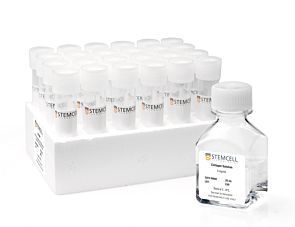
MegaCult™-C Staining Kit for CFU-Mk

Staining kit for human CFU-Mk assays

MegaCult™-C Staining Kit for CFU-Mk

Staining kit for human CFU-Mk assays

From: 3,086 USD
Catalog #
(Select a product)
Staining kit for human CFU-Mk assays
Add to Wish List

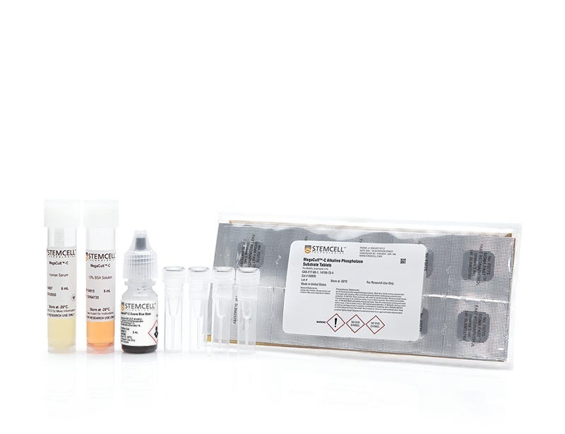
What's Included

  • MegaCult™-C Primary Antibody, 360 µL (Catalog #04803)
  • MegaCult™-C Control Antibody, 100 µL (Catalog #04804)
  • MegaCult™-C Human Serum, 6 mL (Catalog #04807)
  • MegaCult™-C Alkaline Phosphatase Substrate Tablets, 1 pack (Catalog #04809)
  • MegaCult™-C Avidin Alkaline Phosphatase Conjugate, 200 µL (Catalog #04905)
  • MegaCult™-C Biotin-Conjugated Goat Anti-Mouse IgG, 125 µL (Catalog #04906)
  • MegaCult™-C Evans Blue Stain, 5 mL (Catalog #04913)
  • MegaCult™-C 10% BSA, 6 mL (Catalog #04915)

Overview

Stain fixed and dehydrated CFU-Mk colonies cultured in MegaCult™-C collagen-based medium using the reagents included in MegaCult™-C Staining Kit for CFU-Mk.
• MegaCult™-C Primary Antibody
• MegaCult™-C Control Antibody
• MegaCult™-C Human Serum
• MegaCult™-C Alkaline Phosphatase Substrate Tablets
• MegaCult™-C Biotin-Conjugated Goat Anti-Mouse IgG
• MegaCult™-C Evans Blue Stain
• MegaCult™-C 10% BSA

Megakaryocytes and platelets expressing the glycoprotein IIb/IIIa are stained with anti-human CD41 antibody and an alkaline phosphatase detection system. The kit is also suitable for the detection of bipotential erythroid-megakaryocyte progenitor cells (BFU-E/Mk).

For your convenience, the kit components are also available for purchase individually, upon request.
Cell Type
Hematopoietic Stem and Progenitor Cells
Species
Human
Application
Colony Assay, Immunocytochemistry
Brand
MegaCult
Area of Interest
Stem Cell Biology

Data Figures

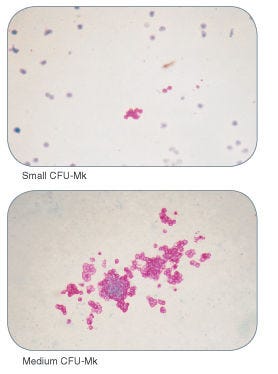
Examples of colonies derived from human megakaryocyte progenitors

Figure 1. Examples of Colonies Derived from Human Megakaryocyte Progenitors

Protocols and Documentation

Find supporting information and directions for use in the Product Information Sheet or explore additional protocols below.

Document Type
Product Name
Catalog #
Lot #
Language
Catalog #
04962
Lot #
All
Language
English
Document Type
Technical Manual
Catalog #
04962
Lot #
All
Language
English
Document Type
Safety Data Sheet 1
Catalog #
04962
Lot #
All
Language
English
Document Type
Safety Data Sheet 2
Catalog #
04962
Lot #
All
Language
English
Document Type
Safety Data Sheet 3
Catalog #
04962
Lot #
All
Language
English
Document Type
Safety Data Sheet 4
Catalog #
04962
Lot #
All
Language
English
Document Type
Safety Data Sheet 5
Catalog #
04962
Lot #
All
Language
English
Document Type
Safety Data Sheet 6
Catalog #
04962
Lot #
All
Language
English
Document Type
Safety Data Sheet 7
Catalog #
04962
Lot #
All
Language
English

Applications

This product is designed for use in the following research area(s) as part of the highlighted workflow stage(s). Explore these workflows to learn more about the other products we offer to support each research area.

Resources and Publications

Frequently Asked Questions

Why is the MegaCult™-C formulation serum free?

MegaCult™-C is formulated without FBS to avoid inhibition of CFU-Mk growth by TGF beta and Platelet Factor-4, which are often present in the serum.

Why use semi-solid media?

Semi-solid media (such as methylcellulose-based or collagen-based) allow the clonal progeny of a single progenitor cell to stay together so you can recognize distinct colonies.

Publications (5)

Comparison of four serum-free, cytokine-free media for analysis of endogenous erythroid colony growth in polycythemia vera and essential thrombocythemia. Dobo I et al. The hematology journal : the official journal of the European Haematology Association / EHA 2001 JAN

Abstract

INTRODUCTION: The assay of endogenous erythroid colony formation (EEC), a characteristic of polycythemia vera and essential thrombocythemia, is not standardized. In this multicentric study, we tested four semisolid, serum-free, cytokine-free media based on either methylcellulose (M1, M2) or collagen (C1, C2) commercialized for the EEC assay. MATERIALS AND METHODS: Bone marrow mononuclear cells (BMMC) from 73 individuals (62 patients with either polycythemia vera (26), essential thrombocythemia (19), secondary polyglobuly (17) or chronic myeloid leukemia (2) and 11 healthy donors) were grown in parallel in the four media without, or with 0.01 U/ml erythropoietin (EPo). RESULTS: In all four media EEC formation was specific, as it was not observed in cultures of patients with secondary polyglobuly or chronic myeloid leukemia, nor of healthy donors. Analysis of fresh or MGG-stained collagen gel cultures allowed detection of EEC formation significantly more frequently than methylcellulose-based media; addition of 0.01 U/ml of EPo had little or no effect on EEC formation. Collagen-based medium C1 gave better results than the other media tested: the 'C1' EEC assay was positive for 68.2% of polycythemia vera cultures with significantly higher median EEC numbers (6.5/10(5) BMMC for patients with one major criteria of polycythemia vera and 19 and 21/10(5) BMMC for patients with two or three major criteria, respectively). Medium C1 was also better for essential thrombocythemia cultures with 47.4% of positive results but with a low median EEC number (6.7/10(5) BMMC). When associated with the ELISA dosage of serum EPo, the 'C1' EEC assay allowed confirmation or elimination of the diagnosis of polycythemia vera for 91% (20/22) of polyglobulic patients. CONCLUSION: We propose that serum-free collagen-based culture systems be considered to standardize the EEC assay, now part of the new criteria of polycythemia vera.
Endogenous erythroid and megakaryocytic colony formation in serum-free, cytokine-free collagen gels. Dobo I et al. Journal of hematotherapy & stem cell research 1999 DEC

Abstract

We studied the suitability of collagen-based semisolid medium for assay of endogenous erythroid colony formation performed in myeloproliferative disorders. Bone marrow (BM) mononuclear cells (MNC) from 103 patients suspected of having polycythemia vera (PV, 76 patients) or essential thrombocythemia (ET, 27 patients) were grown in collagen-based, serum-free, cytokine-free semisolid medium. Colony analysis at day 8 or 10 showed that this collagen assay is specific, as endogenous growth of erythroid colonies was never observed in cultures of 16 healthy donors and 6 chronic myelogenous leukemia (CML) patients. Endogenous erythroid colony formation was observed in 53.3% of patients suspected of PV, with only 15.4% of positive cultures for patients with 1 minor PV criterion and 72% (p = 0.009) of positive cultures for patients with textgreater or =2 minor or 1 major PV criterion. Similarly, endogenous growth of erythroid colonies was found in 44.4% of patients suspected of ET, with 31.6% of positive cultures for patients with 1 ET criterion versus 75% for patients with textgreater or =2 ET criteria. In addition, we found that in collagen gels, tests of erythropoietin (EPO) hypersensitivity in the presence of 0.01 or 0.05 U/ml of EPO and tests of endogenous colony-forming units-megakaryocyte (CFU-MK) formation cannot be used to detect PV or ET, as these tests were positive for, respectively, 21.4% and 50% of healthy donors and 83% and 50% of CML patients. A retrospective analysis suggests that collagen assays are more sensitive than methylcellulose assays to assess endogenous growth of erythroid colonies. In summary, serum-free collagen-based colony assays are simple and reliable assays of endogenous growth of erythroid colonies in myeloproliferative diseases. They also appear to be more sensitive than methylcellulose-based assays.
Quantitation and characterization of human megakaryocyte colony-forming cells using a standardized serum-free agarose assay. Hogge D et al. British journal of haematology 1997 MAR

Abstract

Human progenitors of the megakaryocyte (Mk) lineage were detected by their ability to generate colonies-containing from 3 to textgreater 100 Mk, detectable as glycoprotein IIb/IIIa+ cells in APAAP-stained whole mount agarose cultures. Optimal growth conditions were achieved through the use of a defined serum substitute and a suitable cocktail of recombinant cytokines. Under these culture conditions, the smallest Mk-containing colonies (CFC-Mk) were detectable within a week followed by colonies containing larger numbers of Mk over the ensuing 2 weeks. The total number of CFC-Mk at 18-21 d was linearly related to the number of cells plated. Variation in the cytokines added showed that thrombopoietin (TPO) or IL-3 alone would support the formation of large numbers of CFC-Mk. However, optimal yields of colonies containing cells of both Mk and non-Mk lineages required the addition of other growth factors, of which a combination of IL-3, IL-6, GM-CSF and Steel factor (SF) +/- TPO was the best of those tested. The further addition of erythropoietin to this combination reduced the number of large pure' Mk colonies seen and in their place a corresponding number of mixed erythroid-Mk colonies became detectable. Flt3-ligand alone was unable to support the growth of CFC-Mk nor did it enhance their growth when combined with other factors. Plating of FACS-sorted sub-populations of CD34+ marrow cells in both serum-free agarose and methylcellulose assays demonstrated that most CFC-Mk are generated from CD34+ cells that are CD45RA- and CD71+�