Interested in trying STEMCELL's products for respiratory research? Fill out this form to request information about introductory offers.
Request Pricing
Thank you for your interest in this product.
Please provide us with your contact information and your local representative
will contact you with a customized quote. Where appropriate, they can also assist you with a(n):
Estimated delivery time for your area
Product sample or exclusive offer
In-lab demonstration
By submitting this form, you are providing your consent to STEMCELL Technologies Canada Inc. and its subsidiaries and affiliates (“STEMCELL”) to collect and use your information, and send you newsletters and emails in accordance with our privacy policy. Please contact us with any questions that you may have. You can unsubscribe or change your email preferences at any time.
PneumaCult™-ALI Medium (Catalog #05001) is a serum- and BPE-free medium for the culture of human airway epithelial cells at the air-liquid interface (ALI). Airway epithelial cells cultured in PneumaCult™-ALI Medium undergo extensive mucociliary differentiation to form a pseudostratified epithelium that exhibits morphological and functional characteristics similar to those of the human airway in vivo. PneumaCult™-ALI Medium is also available in a kit that includes 12 mm Transwell® inserts (Catalog #05021) or 6.5 mm Transwell® inserts (Catalog #05022).
Together, PneumaCult™-ALI Medium and PneumaCult™-Ex Plus Medium (Catalog #05040) constitute a fully integrated BPE-free culture system for in vitro human airway modeling that is also compatible with primary human nasal epithelial cells. This robust and defined system is a valuable tool for basic respiratory research, toxicity studies, and drug development.
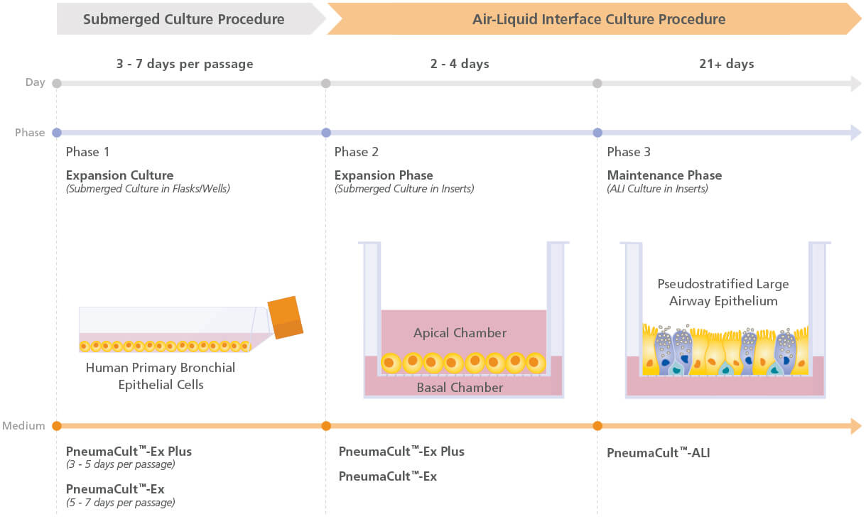
Figure 1. Overview of the PneumaCult™ Culture System
Expansion of human bronchial epithelial cells (HBECs) in submerged culture is performed with PneumaCult™-Ex Plus or PneumaCult™-Ex. During the early Expansion Phase of the ALI culture procedure, PneumaCult™-Ex Plus or PneumaCult™-Ex is applied to the apical and basal chambers. Upon reaching confluence, the culture is air-lifted by removing the culture medium from both chambers, and adding PneumaCult™-ALI to the basal chamber only. Differentiation into a pseudostratified mucociliary epithelium is obtained following 21-28 days of incubation and can be maintained for more than one year.
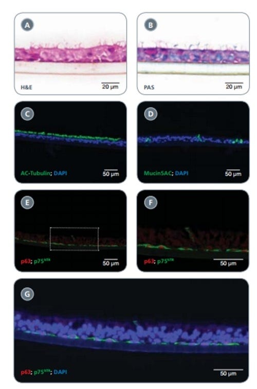
Figure 2. HBECs Cultured in PneumaCult™-Ex Successfully Differentiate into a Pseudostratified Mucociliary Epithelium with PneumaCult™-ALI
Early-passage (P1-3) HBECs cultured in PneumaCult™-Ex successfully differentiate when cultured at air-liquid interface with PneumaCult™-ALI for 28 days. H&E staining revealed the pseudostratifi ed structure of the epithelium with cilia present at the apical surface (A). Periodic acid-Schiff staining demonstrated the presence of goblet cells (B). The presence of ciliated and goblet cells was also demonstrated by immunofl uorescence staining of cilia marker acetylated (AC)-Tubulin (green; C) and the goblet cell marker Mucin5AC (green; D). Appropriate positioning of basal cells along the transwell insert was visualized by immunofl uorescence staining using the basal cell markers p75NTR (green) and p63 (red; E,F). A representative merged image indicates the apical cells, detected by DAPI alone, positioned along the epithelium and in close contact with the basal cells (detected by DAPI, p63 and p75NTR co-labeling) located along the insert (G).
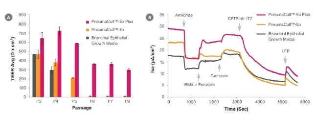
Figure 3. Electrophysiological characterization of differentiated HBECs (P4) that were expanded in PneumaCult™-Ex Plus, PneumaCult™-Ex, and Bronchial Epithelial Growth Media
TEER (A) and representative characterization of the ion channel activities (B) for ALI cultures at 28 days post air-lift using HBECs expanded in PneumaCult™-Ex Plus, PneumaCult™-Ex, or Bronchial Epithelial Growth Media. Amiloride: ENaC inhibitor. IBMX and Forskolin: CFTR activators. Genistein: CFTR potentiator. CFTRinh-172: CFTR inhibitor. UTP: Calciumactivated Chloride channels (CaCCs) activator. All ALI differentiation cultures were performed using PneumaCult™-ALI.
This product is designed for use in the following research area(s) as part
of the highlighted workflow stage(s). Explore these workflows to learn more about the other products we
offer to support each research area.
Choice of Differentiation Media Significantly Impacts Cell Lineage and Response to CFTR Modulators in Fully Differentiated Primary Cultures of Cystic Fibrosis Human Airway Epithelial Cells.
V. Saint-Criq et al.
Cells 2020 sep
Abstract
In vitro cultures of primary human airway epithelial cells (hAECs) grown at air-liquid interface have become a valuable tool to study airway biology under normal and pathologic conditions, and for drug discovery in lung diseases such as cystic fibrosis (CF). An increasing number of different differentiation media, are now available, making comparison of data between studies difficult. Here, we investigated the impact of two common differentiation media on phenotypic, transcriptomic, and physiological features of CF and non-CF epithelia. Cellular architecture and density were strongly impacted by the choice of medium. RNA-sequencing revealed a shift in airway cell lineage; one medium promoting differentiation into club and goblet cells whilst the other enriched the growth of ionocytes and multiciliated cells. Pathway analysis identified differential expression of genes involved in ion and fluid transport. Physiological assays (intracellular/extracellular pH, Ussing chamber) specifically showed that ATP12A and CFTR function were altered, impacting pH and transepithelial ion transport in CF hAECs. Importantly, the two media differentially affected functional responses to CFTR modulators. We argue that the effect of growth conditions should be appropriately determined depending on the scientific question and that our study can act as a guide for choosing the optimal growth medium for specific applications.
Glutaredoxin deficiency promotes activation of the transforming growth factor beta pathway in airway epithelial cells, in association with fibrotic airway remodeling.
S. B. Chia et al.
Redox biology 2020 sep
Abstract
S-glutathionylation of reactive protein cysteines is a post-translational event that plays a critical role in transducing signals from oxidants into biological responses. S-glutathionylation can be reversed by the deglutathionylating enzyme glutaredoxin (GLRX). We have previously demonstrated that ablation of Glrx sensitizes mice to the development of parenchymal lung fibrosis(1). It remains unclear whether GLRX also controls airway fibrosis, a clinical feature relevant to asthma and chronic obstructive pulmonary disease, and whether GLRX controls the biology of airway epithelial cells, which have been implicated in the pathophysiology of these diseases. In the present study we utilized a house dust mite (HDM) model of allergic airway disease in wild type (WT) and Glrx-/- mice on a C57BL/6 background prone to develop airway fibrosis, and tracheal basal stem cells derived from WT mice, global Glrx-/- mice, or bi-transgenic mice allowing conditional ablation of the Glrx gene. Herein we show that absence of Glrx led to enhanced HDM-induced collagen deposition, elevated levels of transforming growth factor beta 1 (TGFB1) in the bronchoalveolar lavage, and resulted in increases in airway hyperresponsiveness. Airway epithelial cells isolated from Glrx-/- mice or following conditional ablation of Glrx showed spontaneous increases in secretion of TGFB1. Glrx-/- basal cells also showed spontaneous TGFB pathway activation, in association with increased expression of mesenchymal genes, including collagen 1a1 and fibronectin. Overall, these findings suggest that GLRX regulates airway fibrosis via a mechanism(s) that involve the plasticity of basal cells, the stem cells of the airways.
Functional rescue of c.3846G\textgreaterA (W1282X) in patient-derived nasal cultures achieved by inhibition of nonsense mediated decay and protein modulators with complementary mechanisms of action.
O. Laselva et al.
Journal of cystic fibrosis : official journal of the European Cystic Fibrosis Society 2020 sep
Abstract
BACKGROUND The nonsense mutation, c.3846G{\textgreater}A (aka: W1282X-CFTR) leads to a truncated transcript that is susceptible to nonsense-mediated decay (NMD) and produces a shorter protein that is unstable and lacks normal channel activity in patient-derived tissues. However, if overexpressed in a heterologous expression system, the truncated mutant protein has been shown to mediate CFTR channel function following the addition of potentiators. In this study, we asked if a quadruple combination of small molecules that together inhibit nonsense mediated decay, stabilize both halves of the mutant protein and potentiate CFTR channel activity could rescue the functional expression of W1282X-CFTR in patient derived nasal cultures. METHODS We identified the CFTR domains stabilized by corrector compounds supplied from AbbVie using a fragment based, biochemical approach. Rescue of the channel function of W1282X.-CFTR protein by NMD inhibition and small molecule protein modulators was studied using a bronchial cell line engineered to express W1282X and in primary nasal epithelial cultures derived from four patients homozygous for this mutation. RESULTS We confirmed previous studies showing that inhibition of NMD using the inhibitor: SMG1i, led to an increased abundance of the shorter transcript in a bronchial cell line. Interestingly, on top of SMG1i, treatment with a combination of two new correctors developed by Galapagos/AbbVie (AC1 and AC2-2, separately targeting either the first or second half of CFTR and promoting assembly, significantly increased the potentiated channel activity by the mutant in the bronchial epithelial cell line and in patient-derived nasal epithelial cultures. The average rescue effect in primary cultures was approximately 50{\%} of the regulated chloride conductance measured in non-CF cultures. CONCLUSIONS These studies provide the first in-vitro evidence in patient derived airway cultures that the functional defects incurred by W1282X, has the potential to be effectively repaired pharmacologically.
Polystyrene plate with lid and polyester membrane inserts for cell culture that feed basolaterally
Item added to your cart
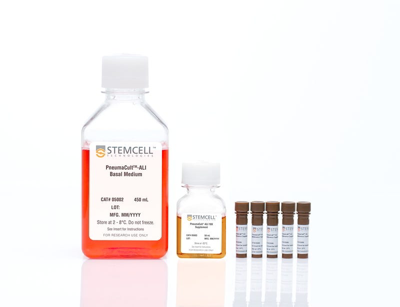
PneumaCult™-ALI Medium
Interested in trying STEMCELL's products for respiratory research? Fill out this form to request information about introductory offers.
Quality Statement:
PRODUCTS ARE FOR RESEARCH USE ONLY AND NOT INTENDED FOR HUMAN OR ANIMAL DIAGNOSTIC OR THERAPEUTIC USES UNLESS OTHERWISE STATED. FOR ADDITIONAL INFORMATION ON QUALITY AT STEMCELL, REFER TO WWW.STEMCELL.COM/COMPLIANCE.